. 2013 May;9(5):e1003352.
doi: 10.1371/journal.ppat.1003352. Epub 2013 May 9.

Gain-of-sensitivity mutations in a Trim5-resistant primary isolate of pathogenic SIV identify two independent conserved determinants of Trim5α specificity

Affiliations

Gain-of-sensitivity mutations in a Trim5-resistant primary isolate of pathogenic SIV identify two independent conserved determinants of Trim5α specificity

Kevin R McCarthy et al. PLoS Pathog. 2013 May.

Abstract

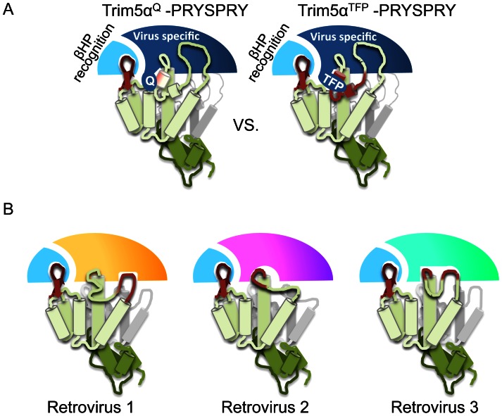
Retroviral capsid recognition by Trim5 blocks productive infection. Rhesus macaques harbor three functionally distinct Trim5 alleles: Trim5α(Q) , Trim5α(TFP) and Trim5(CypA) . Despite the high degree of amino acid identity between Trim5α(Q) and Trim5α(TFP) alleles, the Q/TFP polymorphism results in the differential restriction of some primate lentiviruses, suggesting these alleles differ in how they engage these capsids. Simian immunodeficiency virus of rhesus macaques (SIVmac) evolved to resist all three alleles. Thus, SIVmac provides a unique opportunity to study a virus in the context of the Trim5 repertoire that drove its evolution in vivo. We exploited the evolved rhesus Trim5α resistance of this capsid to identify gain-of-sensitivity mutations that distinguish targets between the Trim5α(Q) and Trim5α(TFP) alleles. While both alleles recognize the capsid surface, Trim5α(Q) and Trim5α(TFP) alleles differed in their ability to restrict a panel of capsid chimeras and single amino acid substitutions. When mapped onto the structure of the SIVmac239 capsid N-terminal domain, single amino acid substitutions affecting both alleles mapped to the β-hairpin. Given that none of the substitutions affected Trim5α(Q) alone, and the fact that the β-hairpin is conserved among retroviral capsids, we propose that the β-hairpin is a molecular pattern widely exploited by Trim5α proteins. Mutations specifically affecting rhesus Trim5α(TFP) (without affecting Trim5α(Q) ) surround a site of conservation unique to primate lentiviruses, overlapping the CPSF6 binding site. We believe targeting this site is an evolutionary innovation driven specifically by the emergence of primate lentiviruses in Africa during the last 12 million years. This modularity in targeting may be a general feature of Trim5 evolution, permitting different regions of the PRYSPRY domain to evolve independent interactions with capsid.


Conflict of interest statement

The authors have declared that no competing interests exist.

Figures

Figure 1
Figure 1. Differential restriction of primate lentiviruses by rhesus Trim5αTFP and Trim5αQ alleles.
GFP reporter viruses were used to infect CRFK cells expressing the rhesus Trim5αTFP allele mamu3 (TFP) and the rhesus Trim5αQ allele mamu4 (Q). Infectivity on empty vector control cells is shown (Ctrl). (A) HIV-1nl4.3. (B) SIVmac239. (C) SIVsmE041. (D) SIVsmE543-3. (E) SIVagmTan-1. (F) HIV-2Rod. Infections were done in triplicate. Error bars indicate SEM. These results are representative of at least 3 independent experiments.
Figure 2
Figure 2. Distribution of amino acid differences between HIV-1nl4.3 and SIVmac239.
(A) Sequence alignment of HIV-1nl4.3 and SIVmac239 CAs. Surface features are indicated on the top, internal α-helices on the bottom. Amino acid differences between the two viruses are in bold type. (B) Structure of the HIV-1nl4.3 N-terminal domain (PDB: 3GV2). The β-hairpin (BHP), the linker connecting the β-hairpin to helix 1 (linker), helix 6 (h6) and 4–5 loop (4–5L) are indicated. Additional α-helices are numbered αH1-αH7. Residues that are identical in HIV-1nl4.3 and SIvamc239 are in red, residues that differ are in blue.
Figure 3
Figure 3. Rhesus Trim5αs recognize the capsid surface.
The indicated GFP reporter viruses were used to infect CRFK cells expressing rhesus Trim5αTFP alleles mamu1, mamu2 and mamu3 (TFP) and the Trim5αQ alleles mamu4 and mamu5 (Q). Infectivity on empty vector control cells is shown (Ctrl). (A) HIV-1nl4.3. (B) SIVmac239. (C) SIV-HIVinterior. (D) SIV-HIVsurface. (E) HIV-SIVsurface. (F) HIV-SIVsurface25. (G) SIV-HIVbhp. (H) SIV-HIVbhpQΔ7.(I) SIV-HIV4–5L. (J) SIV-HIVh6. Infections were done in triplicate. Error bars indicate SEM. These results are representative of at least 3 independent experiments.
Figure 4
Figure 4. Structure of the SIVmac239 Capsid N-terminal domain.
(A) Structure of the SIVmac239 CA N-terminal domain at 2.9 Å resolution. There was no clear density for Pro88, and thus, it was omitted from the structure. A dashed line is used to indicate its place. (B) Comparison of the SIVmac239 β-hairpin and 4–5 loop to all other wild type HIV-1 and HIV-2 X-ray structures deposited in the PDB. HIV-1 structures are colored dark gray, except PDB: 2X2D, which is colored red (and used in all subsequent comparisons). HIV-2 structures are colored light gray, and the SIVmac239 N-terminal domain is colored blue. (C and D) Locations of amino acids mutations associated with rhesus Trim5αQ (C) and rhesus Trim5αTFP (D) restriction from Table 1. Blue spheres indicate amino acid differences that do not impact Trim5α restriction. Orange spheres show the location of mutations associated with 2.5–5 fold gains in sensitivity to rhesus Trim5α relative to SIVmac239. Red spheres indicate positions associated with >5 fold gains in sensitivity to rhesus Trim5α. Images created in PyMol.
Figure 5
Figure 5. Mutations modulating rhesus Trim5αTFP restriction ring a conserved surface patch.
(A) Top row: Orientations of the SIVmac239 capsid used for Figure 5A. Middle row: Surface representation of the SIVmac239 capsid N-terminal domain colored to reflect amino acid conservation across divergent primate lentiviruses. The number of unique amino acids found at each position in an amino acid alignment of eleven divergent primate lentiviruses (Figure S7) was scored and colored according to the legend: Orange ≥4 unique amino acids at the specified position, yellow 3 unique amino acids at the specified position, light gray 2 unique residues at the specified position and dark gray 1 amino acid (100% conservation) at the specified position. The location of the conserved surface patch is indicated by dashed lines. Bottom panel: Locations of mutations that are associated with a >2.5 fold gain in sensitivity to rhTrim5αTFP are shown in dark red. (B) Atomic view of the conserved surface patch. For reference the SIVmac239 and HIV-1 (2X2D) ribbon diagrams are shown in light blue and pink, respectively. The amino acids that make up the conserved surface patch are shown in sticks that are colored according to the capsid ribbon diagram, SIVmac239 in light blue and HIV-1 in light red. Variable positions shown to modulate rhesus Trim5αTFP sensitivity are colored in dark blue (SIVmac239) and dark red sticks (HIV-1) for emphasis. Images created in PyMol.
Figure 6
Figure 6. Evolutionary origins of the Trim5αTFP allele.
A Cladogram depicting the evolutionary relationships among Trim5 coding sequences from 16 extant primate species. Major divergence times are in bold, approximate dates of events discussed in the text are indicated with arrows. For each species/allele, the amino acid sequence corresponding to residues 335–346 (relative to rhesus Trim5) is shown; species names followed by numbers indicate multiple alleles. Residues with dN/dS >1 and a high posterior probability of positive selection are indicated by † (posterior probability >99%) or * (posterior probability >95%).
Figure 7
Figure 7. A Proposed model for the evolution of novel Trim5α variants.
The rhTrim5αQ alleles and the rhTrim5αTFP alleles share the ability to recognize lentiviral β-hairpins. The rhTrim5αTFP alleles evolved to recognize the conserved surface patch. We believe this observation underscores an inherent uncoupling between capsid recognition modules within the PRYSPRY domain. The β-hairpin is a conserved feature found in all reported retroviral capsid structures, and therefore a convenient target for host proteins that evolve to recognize a broad range of retroviruses. We believe β-hairpin targeting is a conserved feature of Trim5α proteins and allows for the evolution of specificity of capsid recognition. (A) Evolution of conserved surface patch recognition. The rhTrim5αQ allele is capable of strongly recognizing the β-hairpin (dark red) and able to engage in a weaker contact (pink) with helix 6, at one edge of the conserved surface patch. Recognition of these two features is conserved between rhTrim5αQ and rhTrim5αTFP alleles and therefore unaffected by the Q/TFP polymorphism. The region of the PRYSPRY that encodes for the intrinsic β-hairpin recognition module is colored light blue and the module that can adapt to specific viruses is colored dark blue. We propose that the polymorphic region of variable loop 1 (V1) is uncoupled from intrinsic β-hairpin recognition (by of another region within the PRYSPRY) allowing it to tolerate mutations such as the 6 nucleotide insertion. In this model, the rhTrim5αTFP allele engages in similar contacts as the rhTrim5αQ allele, but has gained the ability to target the conserved surface patch (dark red). (B) The ability to recognize a conserved retroviral element, even if it allows very weak associations retroviral capsids, can allow for the selection of additional capsid binding modules within the PRYSPRY domain allowing it to adapt to specific retroviral pressures. Hypothetical adaptation to different retroviral targets are depicted as differently colored PRYSPRY domains. Together this process could lead to the breadth and specificity observed among Trim5α orthologs. For simplicity this model is depicted with one PRYSPRY domain recognizing one capsid monomer, although the stoichiometry or orientation of Trim5α binding is not known at this time.

Similar articles

Cited by

References

    1. Stremlau M, Owens CM, Perron MJ, Kiessling M, Autissier P, et al. (2004) The cytoplasmic body component TRIM5alpha restricts HIV-1 infection in Old World monkeys. Nature 427: 848–853. - PubMed
    1. Newman RM, Johnson WE (2007) A brief history of TRIM5alpha. AIDS Rev 9: 114–125. - PubMed
    1. Schaller T, Hue S, Towers GJ (2007) An active TRIM5 protein in rabbits indicates a common antiviral ancestor for mammalian TRIM5 proteins. J Virol 81: 11713–11721. - PMC - PubMed
    1. Ylinen LM, Keckesova Z, Webb BL, Gifford RJ, Smith TP, et al. (2006) Isolation of an active Lv1 gene from cattle indicates that tripartite motif protein-mediated innate immunity to retroviral infection is widespread among mammals. J Virol 80: 7332–7338. - PMC - PubMed
    1. Rahm N, Yap M, Snoeck J, Zoete V, Munoz M, et al. (2011) Unique spectrum of activity of prosimian TRIM5alpha against exogenous and endogenous retroviruses. J Virol 85: 4173–4183. - PMC - PubMed

Publication types

Substances

LinkOut - more resources