. 2013 May;9(5):e1003368.
doi: 10.1371/journal.ppat.1003368. Epub 2013 May 9.

T-Cell tropism of simian varicella virus during primary infection

Affiliations

T-Cell tropism of simian varicella virus during primary infection

Werner J D Ouwendijk et al. PLoS Pathog. 2013 May.

Abstract

Varicella-zoster virus (VZV) causes varicella, establishes a life-long latent infection of ganglia and reactivates to cause herpes zoster. The cell types that transport VZV from the respiratory tract to skin and ganglia during primary infection are unknown. Clinical, pathological, virological and immunological features of simian varicella virus (SVV) infection of non-human primates parallel those of primary VZV infection in humans. To identify the host cell types involved in virus dissemination and pathology, we infected African green monkeys intratracheally with recombinant SVV expressing enhanced green fluorescent protein (SVV-EGFP) and with wild-type SVV (SVV-wt) as a control. The SVV-infected cell types and virus kinetics were determined by flow cytometry and immunohistochemistry, and virus culture and SVV-specific real-time PCR, respectively. All monkeys developed fever and skin rash. Except for pneumonitis, pathology produced by SVV-EGFP was less compared to SVV-wt. In lungs, SVV infected alveolar myeloid cells and T-cells. During viremia the virus preferentially infected memory T-cells, initially central memory T-cells and subsequently effector memory T-cells. In early non-vesicular stages of varicella, SVV was seen mainly in perivascular skin infiltrates composed of macrophages, dendritic cells, dendrocytes and memory T-cells, implicating hematogenous spread. In ganglia, SVV was found primarily in neurons and occasionally in memory T-cells adjacent to neurons. In conclusion, the data suggest the role of memory T-cells in disseminating SVV to its target organs during primary infection of its natural and immunocompetent host.


Conflict of interest statement

Co-author ADMEO wishes to declare, for the avoidance of any misunderstanding on competing interests, that he co-founded and is chief scientific officer of Viroclinics Biosciences, a company set up in collaboration with Erasmus MC. However, for clarification, no materials or support were received from the company, and no agreements were in place concerning the execution or publication of this work. This does not alter our adherence to all PLoS Pathogens policies on sharing data and materials. The authors have no additional financial interests.

Figures
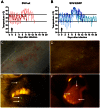
Figure 1
Figure 1. Experimental SVV infection of African green monkeys results in transient fever and skin rash.
(A, B) Fluctuations in body temperature after infection with SVV-wt and SVV-EGFP, respectively, were measured by intraperitoneally implanted temperature transponders during primary infection. Arrows indicate time of SVV inoculation; horizontal lines indicate normal range in body temperature before infection. (C) Vesicular skin rash at 8 dpi with SVV-wt. (D) Macroscopic detection of EGFP fluorescence on skin at 8 dpi with SVV-EGFP. (E) Macroscopic detection of EGFP fluorescence (arrows) on lips at 9 dpi with SVV-EGFP. (F) Macroscopic detection of EGFP-positive lesions (arrows) on tongue at 8 dpi with SVV-EGFP.
Figure 2
Figure 2. Macroscopic and microscopic detection of SVV-infected cells in lungs of infected African green monkeys.
(A) Macroscopic appearance of consolidated dark-red lesions (black arrow) in the lung of an SVV-wt−infected monkey at 13 dpi. (B) Macroscopic detection of EGFP fluorescence in affected area of lung (white arrow) of an SVV-EGFP-infected monkey at 9 dpi. (C) Magnification of the affected area in panel B shows EGFP fluorescence. (D–G) Serial lung sections obtained from an SVV-EGFP−infected monkey at 9 dpi analyzed by immunohistochemistry (IHC) for SVV antigens (D) or by immunofluorescence (IF) for EGFP (E), with two sections analyzed by IHC (F) or IF (G) using normal rabbit serum (NRS) and isotype control antibodies, respectively. Lung sections obtained from an SVV-wt−infected monkey at 9 dpi were analyzed using dual IF for SVV (green) and: cytokeratin (red) (H), CD3 (red) (I), CD68 (red) (J), and CD11c (red) (K) antigens. Arrows indicate double-positive cells. Asterisks indicate autofluorescent erythrocytes. Dashed lines indicate alveolar septa. Br: bronchus. Nuclei were counterstained with DAPI. D–G: 100× magnification; H, J: 400× magnification; I, K: 400× magnification and 2× digital zoom.
Figure 3
Figure 3. SVV preferentially infects myeloid cells and T-cells in lungs of infected African green monkeys.
(A) Bronchoalveolar lavage (BAL) cells obtained at 5, 9 and 13 dpi were analyzed for viral DNA by SVV-specific real-time qPCR. Data are expressed as genome equivalent copies (geq) per 105 BAL cells. (B) BAL cells were analyzed for infectious virus by co-cultivation with BSC-1 cells. (C–F) Percentage of EGFP-positive cells as assessed by flow cytometry in: all BAL cells (C); leukocytes (CD45pos cells), non-leukocytes (CD45neg cells) from BAL samples (D) and leukocyte subsets within BAL (E). Leukocyte subsets were identified based on the differential expression of the following markers: AM/DC = CD45posCD3negCD20negMHC-IIposCD14pos/dim, T-cells = CD45posCD3pos, B-cells = CD45posCD20posMHC-IIpos; and the indicated T-cell subsets (F). AM/DC are BAL-derived lymphocytes expressing markers shared by dendritic cells (DC) and alveolar macrophages (AM). Horizontal bars indicate median values. (D–F) BAL cells were obtained at 5 dpi and data are given as means ± SEM.
Figure 4
Figure 4. SVV infects predominantly memory T-cells in blood after infection in African green monkeys.
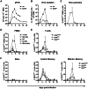
(A) Average SVV DNA load in PBMC of SVV-wt− (closed squares) and SVV-EGFP− (open squares) infected monkeys determined by SVV-specific real-time qPCR. (B) PBMC from SVV-wt− (closed squares) and SVV-EGFP− (open squares) infected monkeys were analyzed for infectious SVV by co-cultivation with BSC-1 cells. (C) PBMC from SVV-EGFP−infected monkeys were analyzed for EGFP expression by flow cytometry. (D) EGFP expression in PBMC subsets from SVV-EGFP−infected monkeys. Data are given as percentage of EGFPpos cells within each lymphocyte subset relative to the total number of PBMC, as determined by flow cytometry. Lymphocyte subsets were defined by differential expression of the following markers: T-cells = CD3posCD16neg cells, B-cells = CD20posMHC-IIposcells, natural killer (NK) cells = CD3negCD16pos cells, dendritic cells (DC) = CD3negCD14negCD16negCD20negCD14negMHC-IIpos cells, and monocytes = CD3negCD14posMHC-IIpos cells. (E and F) Percentage of EGFPpos cells among each T-cell subset relative to the number of CD8bright, CD8dim and CD4pos T-cells (E) and in naive, central memory and effector memory T-cells (F) from SVV-EGFP−infected monkeys as evaluated by flow cytometry. In all panels, data are means ± SEM.
Figure 5
Figure 5. Detection of SVV in lymphoid organs from infected African green monkeys.
(A) Real-time qPCR analysis of SVV DNA load in tonsil, lymph nodes and spleen from SVV-wt− (closed squares) and SVV-EGFP− (open squares) infected monkeys at 9, 13 and 20 dpi. Squares indicate individual tissues, i.e., tonsils (red), tracheobronchial lymph nodes (LN) (green), axillary LN (pink), mandibular LN (blue), inguinal LN (orange) and spleen (black). Horizontal bar indicates the median value. (B–D) Serial sections of tonsil from an SVV-wt−infected monkey stained with hematoxylin and eosin (inset shows a Cowdry type A intranuclear inclusion body) (B) or examined immunohistochemically using rabbit anti-SVV antibodies (C) or control normal rabbit serum (D). Magnification: 200×. The area of tonsils containing multiple intranuclear inclusion bodies contained numerous cells expressing SVV protein. **p<0.01 by Mann-Whitney test.
Figure 6
Figure 6. Detection of SVV-infected cells in varicella skin lesions from infected African green monkeys.
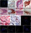
(A, B) Consecutive sections of skin obtained from an SVV-EGFP-infected monkey at 9 dpi and stained by immunofluorescence (IF) for EGFP (A) and by immunohistochemistry (IHC) for SVV antigens (B) show co-localization of SVV proteins and EGFP. Squares indicate the same area of tissue. (C–H) Consecutive sections of skin obtained from an SVV-wt−infected animal at 9 dpi and examined by staining with hematoxylin and eosin (H&E) or by IHC for SVV show virus-induced histopathology and viral proteins in epidermal blisters (C and D), dermal hair follicles (E and F) and dermal sebaceous glands (G and H). (I, J) Consecutive skin sections obtained from an SVV-EGFP-infected monkey at 9 dpi and stained with H&E (I) or by IHC for SVV antigens (J) show blood vessels (asterisks) surrounded by SVV protein-positive cells (arrows). Inset: magnification of the epidermis showing Cowdry type A intranuclear inclusion bodies in panel I (arrowheads) and SVV protein-positive cells in panel J (arrows). (K) Skin section from an SVV-EGFP-infected animal obtained at 9 dpi and double-stained for EGFP (green) and alpha-smooth muscle actin (SMA; red). Asterisks indicate SMA-positive sweat glands, arrowheads indicate SMA-positive blood vessels, and arrows indicate EGFP-positive cells. (L–N) Skin sections obtained at 9 dpi and double-stained for EGFP (green) and: CD68 (red) (L); CD11c (red) (M); and CD3 (red) (N). Arrows indicate dual-stained cells. (O) Skin section obtained at 9 dpi and double-stained for SVV (green) and CD3 (red). Arrows indicate dual-stained cells. A, B: 100× magnification; C–K: 200× magnification; L–O: 400× magnification and 2× digital zoom.
Figure 7
Figure 7. Detection of SVV-infected cells in ganglia of infected African green monkeys.
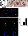
(A) Virus DNA load was determined in ganglia at 9, 13 and 20 dpi by SVV-specific real-time qPCR. Filled and open squares represent pooled ganglia from the same level of the neuraxis from animals infected with SVV-wt and SVV-EGFP, respectively. Colors indicate level of the neuraxis: trigeminal (black), cervical (red), thoracic (blue), lumbar (green) and sacral (pink) ganglia. Horizontal bars represent mean viral DNA load per animal. (B) Immunohistochemical detection of SVV proteins (arrowheads) in a cervical ganglion at 9 dpi. Squares indicate corresponding tissue areas shown at higher magnification in (C) and (D). (E) Dual-immunofluorescence (IF) staining of a thoracic ganglion at 9 dpi for SVV proteins (green) and neural cell adhesion molecule (NCAM; red). Arrows indicate SVV-positive neurons. (F) Dual-IF staining of a thoracic ganglion at 9 dpi for SVV protein (green) and glial fibrillary acidic protein (GFAP; red). Arrow indicates neuron-adjacent SVV-positive cell. (G) Dual-IF staining of a thoracic ganglion from a monkey at 9 dpi for SVV protein (green) and CD3 (red). Arrows indicate SVV-positive T-cells. Asterisks indicate autofluorescent lipofuscin and the borders of the neuronal cell bodies are indicated with dashed lines. (H) Ganglion-derived single-cell suspensions were analyzed by flow cytometry and T-cells were categorized as naive, central memory (CM) and effector memory (EM) T-cells. E–G: nuclei were counterstained with DAPI (blue). ** p<0.01 by Mann-Whitney test. B: 200× magnification; C, D: 400× magnification, 2× digital zoom; E: 400× magnification; F, G: 400× magnification, 2× digital zoom.
Figure 8
Figure 8. Schematic presentation of primary SVV infection.
Figure shows the kinetics of SVV infection and virus-infected cell types in African green monkeys during primary SVV infection. Horizontal lines indicate the time-frame covered by the sampling days. Width of the black bars indicates onset and severity of clinical signs, amount of SVV DNA detected in blood and the sampled organs, and the frequency of SVV-infected cells in peripheral blood during primary SVV infection. Note that BAL samples were obtained no earlier than 5 dpi and animals were euthanized no earlier than 9 dpi. BAL: bronchoalveolar lavage; NK cells: natural killer cells; DC: dendritic cell; TCM: central memory T-cells; TEM: effector memory T-cells.
Figure 9
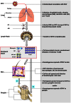
Figure 9. Model of the pathogenesis of primary SVV infection.
Upon intratracheal inoculation of African green monkeys, SVV replicates in the lower respiratory tract and infects lung epithelial cells, alveolar macrophages (AM), dendritic cells (DC) and T-cells. SVV-infected AM and DC may transport the virus to draining lymph nodes and subsequently transfer SVV to local lymphocytes resulting in a cell-associated viremia. Memory T-cells are the predominant SVV-infected lymphocyte subset during viremia and may play a central role in dissemination of SVV to its target organs. SVV reaches the skin by the hematogenous route, presumable via virus-infected memory T-cells, which results in the infection of perivascular macrophages, DC and dendrocytes. Subsequently, SVV may infect epidermal and hair follicle keratinocytes via cell-to-cell spread and cause vesicular skin lesions. SVV may enter ganglia by (a) retrograde axonal transport and/or (b) by viremic spread via virus-infected lymphocytes.

Similar articles

Cited by

References

    1. Cohen JI, Straus SE, Arvin AM (2007) Varicella-zoster virus replication, pathogenesis, and management. In: Knipe DM, Howley PM, editors. Fields Virology. 5th edition. Philadelphia, PA: Lippincott-Williams and Wilkins. pp. 2773–2818.
    1. Grose C (1981) Variation on a theme by Fenner: the pathogenesis of chickenpox. Pediatrics 68: 735–737. - PubMed
    1. Heininger U, Seward JF (2006) Varicella. Lancet 368: 1365–1376. - PubMed
    1. Asano Y, Itakura N, Hiroishi Y, Hirose S, Ozaki T, et al. (1985) Viral replication and immunologic responses in children naturally infected with varicella-zoster virus and in varicella vaccine recipients. J Infect Dis 152: 863–868. - PubMed
    1. Koropchak CM, Graham G, Palmer J, Winsberg M, Ting SF, et al. (1991) Investigation of varicella-zoster virus infection by polymerase chain reaction in the immunocompetent host with acute varicella. J Infect Dis 163: 1016–1022. - PubMed

Publication types