

Liver x receptors protect from development of prostatic intra-epithelial neoplasia in mice
- PMID: 23675307
- PMCID: PMC3649972
- DOI: 10.1371/journal.pgen.1003483
Liver x receptors protect from development of prostatic intra-epithelial neoplasia in mice
Abstract
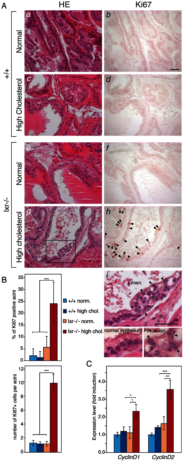
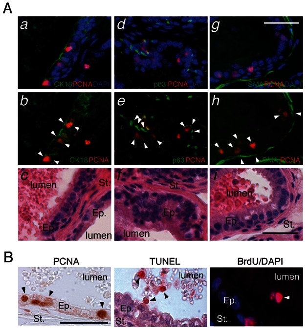
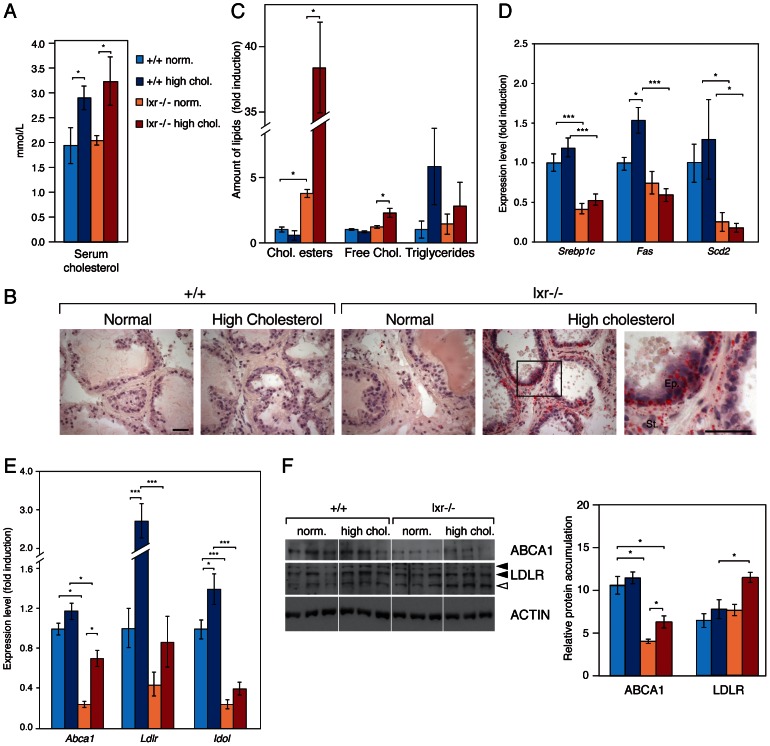
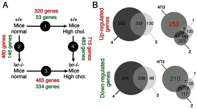
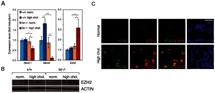
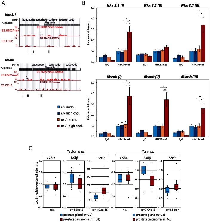
LXR (Liver X Receptors) act as "sensor" proteins that regulate cholesterol uptake, storage, and efflux. LXR signaling is known to influence proliferation of different cell types including human prostatic carcinoma (PCa) cell lines. This study shows that deletion of LXR in mouse fed a high-cholesterol diet recapitulates initial steps of PCa development. Elevation of circulating cholesterol in Lxrαβ-/- double knockout mice results in aberrant cholesterol ester accumulation and prostatic intra-epithelial neoplasia. This phenotype is linked to increased expression of the histone methyl transferase EZH2 (Enhancer of Zeste Homolog 2), which results in the down-regulation of the tumor suppressors Msmb and Nkx3.1 through increased methylation of lysine 27 of histone H3 (H3K27) on their promoter regions. Altogether, our data provide a novel link between LXR, cholesterol homeostasis, and epigenetic control of tumor suppressor gene expression.
Conflict of interest statement
The authors have declared that no competing interests exist.
Figures
Similar articles
-
Down-regulation of human DAB2IP gene expression mediated by polycomb Ezh2 complex and histone deacetylase in prostate cancer.J Biol Chem. 2005 Jun 10;280(23):22437-44. doi: 10.1074/jbc.M501379200. Epub 2005 Apr 6. J Biol Chem. 2005. PMID: 15817459
-
The gene encoding the prostatic tumor suppressor PSP94 is a target for repression by the Polycomb group protein EZH2.Oncogene. 2007 Jul 5;26(31):4590-5. doi: 10.1038/sj.onc.1210248. Epub 2007 Jan 22. Oncogene. 2007. PMID: 17237810
-
BRCA1 and EZH2 cooperate in regulation of prostate cancer stem cell phenotype.Int J Cancer. 2019 Dec 1;145(11):2974-2985. doi: 10.1002/ijc.32323. Epub 2019 May 10. Int J Cancer. 2019. PMID: 30968962
-
Lipids, LXRs and prostate cancer: are HDACs a new link?Biochem Pharmacol. 2013 Jul 1;86(1):168-74. doi: 10.1016/j.bcp.2013.04.005. Epub 2013 Apr 23. Biochem Pharmacol. 2013. PMID: 23618920 Review.
-
EZH2 methyltransferase and H3K27 methylation in breast cancer.Int J Biol Sci. 2012;8(1):59-65. doi: 10.7150/ijbs.8.59. Epub 2011 Nov 18. Int J Biol Sci. 2012. PMID: 22211105 Free PMC article. Review.
Cited by
-
LXR/RXR pathway signaling associated with triple-negative breast cancer in African American women.Breast Cancer (Dove Med Press). 2018 Dec 20;11:1-12. doi: 10.2147/BCTT.S185960. eCollection 2019. Breast Cancer (Dove Med Press). 2018. PMID: 30588086 Free PMC article.
-
The Role of Nuclear Receptors in Prostate Cancer.Cells. 2019 Jun 17;8(6):602. doi: 10.3390/cells8060602. Cells. 2019. PMID: 31212954 Free PMC article. Review.
-
EZH2 is overexpressed in adrenocortical carcinoma and is associated with disease progression.Hum Mol Genet. 2016 Jul 1;25(13):2789-2800. doi: 10.1093/hmg/ddw136. Epub 2016 May 5. Hum Mol Genet. 2016. PMID: 27149985 Free PMC article.
-
Liver X Receptors Enhance Epithelial to Mesenchymal Transition in Metastatic Prostate Cancer Cells.Cancers (Basel). 2024 Aug 6;16(16):2776. doi: 10.3390/cancers16162776. Cancers (Basel). 2024. PMID: 39199549 Free PMC article.
-
Nonalcoholic fatty liver disease: molecular pathways and therapeutic strategies.Lipids Health Dis. 2013 Nov 9;12:171. doi: 10.1186/1476-511X-12-171. Lipids Health Dis. 2013. PMID: 24209497 Free PMC article. Review.
References
-
- Tontonoz P, Mangelsdorf DJ (2003) Liver X receptor signaling pathways in cardiovascular disease. Mol Endocrinol 17: 985–993. - PubMed
-
- Mangelsdorf DJ, Evans RM (1995) The RXR heterodimers and orphan receptors. Cell 83: 841–850. - PubMed
-
- Viennois E, Pommier AJ, Mouzat K, Oumeddour A, El Hajjaji FZ, et al. (2011) Targeting liver X receptors in human health: deadlock or promising trail? Expert Opin Ther Targets 15: 219–232 doi:10.1517/14728222.2011.547853. - DOI - PubMed
-
- Viennois E, Esposito T, Dufour J, Pommier A, Fabre S, et al. (2012) Lxrá regulates the androgen response in prostate epithelium. Endocrinology 153: 3211–3223 doi:10.1210/en.2011-1996. - DOI - PubMed
Publication types
MeSH terms
Substances
LinkOut - more resources
Full Text Sources
Other Literature Sources
Medical
Molecular Biology Databases
