

Postnatal development of cerebellar zones revealed by neurofilament heavy chain protein expression
- PMID: 23675325
- PMCID: PMC3648691
- DOI: 10.3389/fnana.2013.00009
Postnatal development of cerebellar zones revealed by neurofilament heavy chain protein expression
Abstract
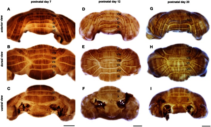
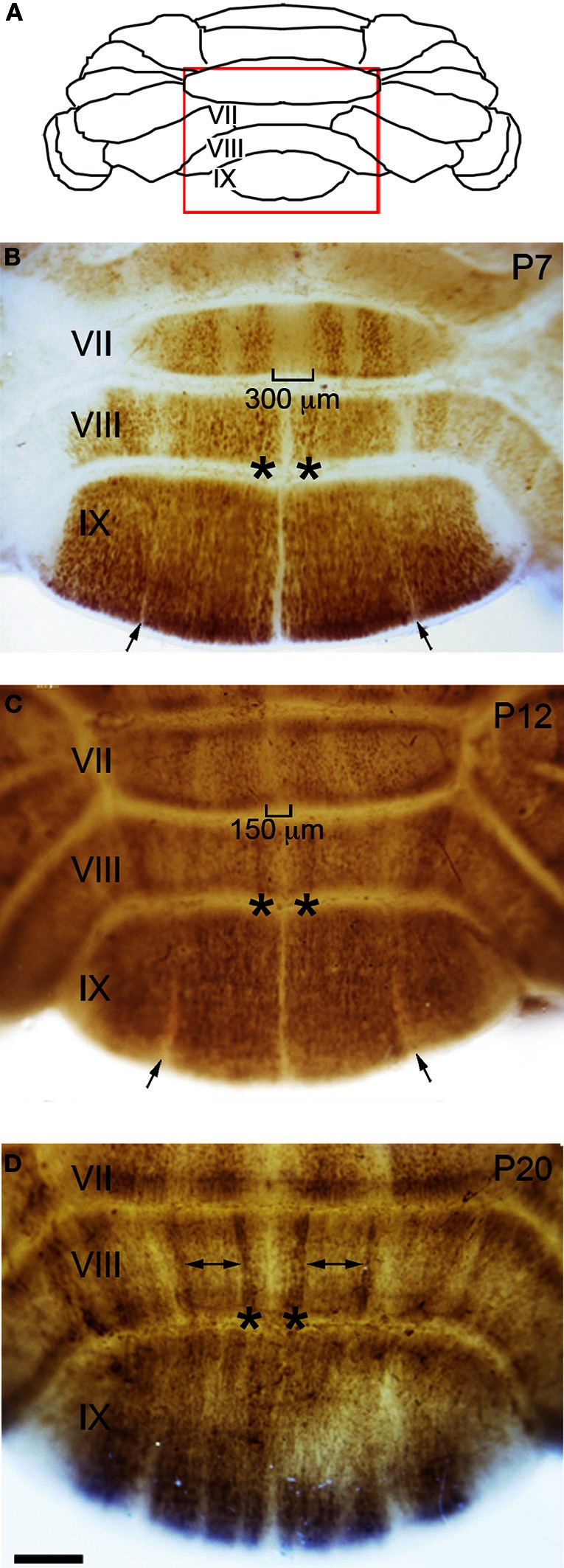
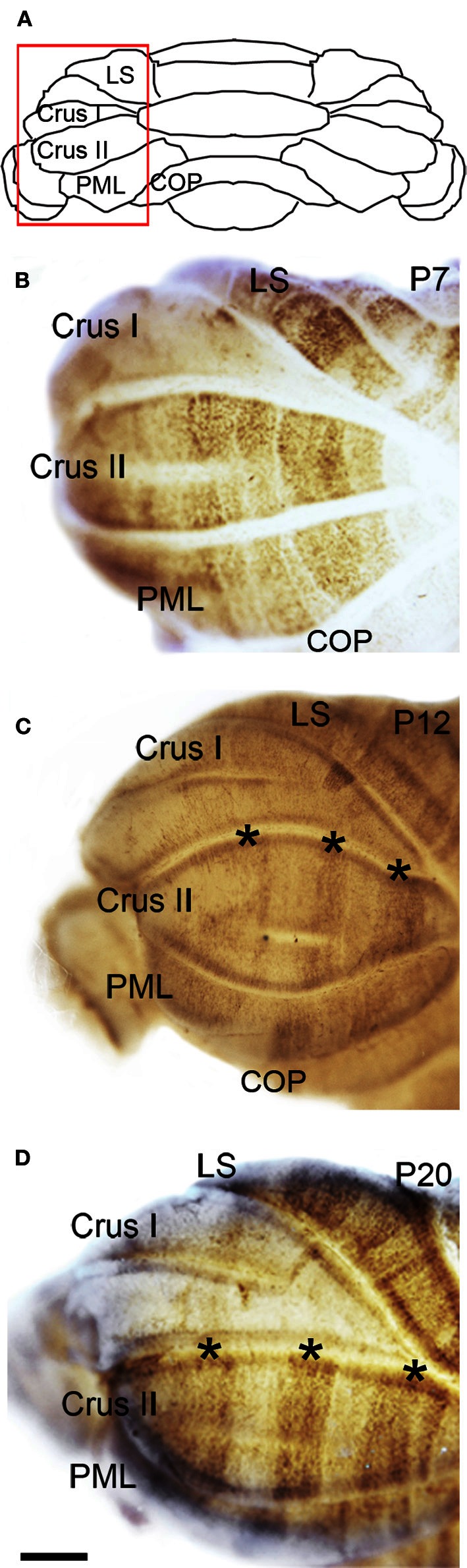
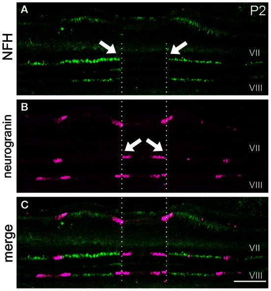
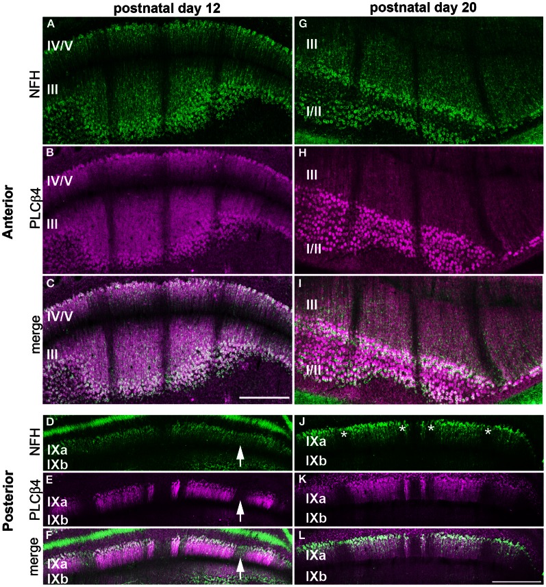
The cerebellum is organized into parasagittal zones that control sensory-motor behavior. Although the architecture of adult zones is well understood, very little is known about how zones emerge during development. Understanding the process of zone formation is an essential step toward unraveling how circuits are constructed to support specific behaviors. Therefore, we focused this study on postnatal development to determine the spatial and temporal changes that establish zonal patterns during circuit formation. We used a combination of wholemount and tissue section immunohistochemistry in mice to show that the cytoskeletal protein neurofilament heavy chain (NFH) is a robust marker for postnatal cerebellar zonal patterning. The patterned expression of NFH is initiated shortly after birth, and compared to the domains of several known zonal markers such as zebrin II, HSP25, neurogranin, and phospholipase Cβ4 (PLCβ4), NFH does not exhibit transient expression patterns that are typically remodeled between stages, and the adult zones do not emerge after a period of uniform expression in all lobules. Instead, we found that throughout postnatal development NFH gradually reveals distinct zones in each cerebellar lobule. The boundaries of individual NFH zones sharpen over time, as zones are refined during the second and third weeks after birth. Double labeling with neurogranin and PLCβ4 further revealed that although the postnatal expression of NFH is spatially and temporally unique, its pattern of zones respects a fundamental and well-known molecular topography in the cerebellum. The dynamics of NFH expression support the hypothesis that adult circuits are derived from an embryonic map that is refined into zones during the first 3-weeks of life.
Keywords: circuit; development; patterning; purkinje cells; topography.
Figures
Similar articles
-
Mossy Fibers Terminate Directly Within Purkinje Cell Zones During Mouse Development.Cerebellum. 2016 Feb;15(1):14-17. doi: 10.1007/s12311-015-0712-6. Cerebellum. 2016. PMID: 26255945 Free PMC article.
-
Neurofilament heavy chain expression reveals a unique parasagittal stripe topography in the mouse cerebellum.Cerebellum. 2011 Sep;10(3):409-21. doi: 10.1007/s12311-010-0156-y. Cerebellum. 2011. PMID: 20127431
-
Phospholipase Cbeta4 expression reveals the continuity of cerebellar topography through development.J Comp Neurol. 2007 Jun 10;502(5):857-71. doi: 10.1002/cne.21352. J Comp Neurol. 2007. PMID: 17436294
-
Insights into cerebellar development and connectivity.Neurosci Lett. 2019 Jan 1;688:2-13. doi: 10.1016/j.neulet.2018.05.013. Epub 2018 May 7. Neurosci Lett. 2019. PMID: 29746896 Free PMC article. Review.
-
The Relationship Between Zebrin Expression and Cerebellar Functions: Insights From Neuroimaging Studies.Front Neurol. 2020 Apr 22;11:315. doi: 10.3389/fneur.2020.00315. eCollection 2020. Front Neurol. 2020. PMID: 32390933 Free PMC article. Review.
Cited by
-
Axonal pathology differentially affects human Purkinje cell subpopulations in the essential tremor cerebellum.Proc Natl Acad Sci U S A. 2025 Jul 8;122(27):e2502024122. doi: 10.1073/pnas.2502024122. Epub 2025 Jun 30. Proc Natl Acad Sci U S A. 2025. PMID: 40587795
-
Mossy Fibers Terminate Directly Within Purkinje Cell Zones During Mouse Development.Cerebellum. 2016 Feb;15(1):14-17. doi: 10.1007/s12311-015-0712-6. Cerebellum. 2016. PMID: 26255945 Free PMC article.
-
Consensus Paper: Cerebellar Development.Cerebellum. 2016 Dec;15(6):789-828. doi: 10.1007/s12311-015-0724-2. Cerebellum. 2016. PMID: 26439486 Free PMC article. Review.
-
Purkinje cell neurotransmission patterns cerebellar basket cells into zonal modules defined by distinct pinceau sizes.Elife. 2020 Sep 29;9:e55569. doi: 10.7554/eLife.55569. Elife. 2020. PMID: 32990595 Free PMC article.
-
Purkinje Cell Patterning-Insights from Single-Cell Sequencing.Cells. 2022 Sep 18;11(18):2918. doi: 10.3390/cells11182918. Cells. 2022. PMID: 36139493 Free PMC article. Review.
References
-
- Ahn A., Dziennis S., Hawkes R., Herrup K. (1994). The cloning of zebrin II reveals its identity with aldolase C. Development 120, 2081–2090 - PubMed
-
- Altman J., Bayer S. (1997). Development of the Cerebellar System: in Relation to its Evolution, Structure, and Functions. Boca Raton, FL: CRC Press
-
- Armstrong C. L., Hawkes R. (2000). Pattern formation in the cerebellar cortex. Biochem. Cell Biol. 78, 551–562 - PubMed
-
- Armstrong C. L., Krueger-Naug A. M., Currie R. W., Hawkes R. (2000). Constitutive expression of the 25-kDa heat shock protein Hsp25 reveals novel parasagittal bands of purkinje cells in the adult mouse cerebellar cortex. J. Comp. Neurol. 416, 383–397 10.1002/(SICI)1096-9861(20000117)416:3<383::AID-CNE9>3.0.CO;2-M - DOI - PubMed
Grants and funding
LinkOut - more resources
Full Text Sources
Other Literature Sources
Research Materials