Activation of PAC1 Receptors in Rat Cerebellar Granule Cells Stimulates Both Calcium Mobilization from Intracellular Stores and Calcium Influx through N-Type Calcium Channels
- PMID: 23675369
- PMCID: PMC3650316
- DOI: 10.3389/fendo.2013.00056
Activation of PAC1 Receptors in Rat Cerebellar Granule Cells Stimulates Both Calcium Mobilization from Intracellular Stores and Calcium Influx through N-Type Calcium Channels
Abstract
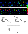
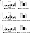
High concentrations of pituitary adenylate cyclase-activating polypeptide (PACAP) and a high density of PACAP binding sites have been detected in the developing rat cerebellum. In particular, PACAP receptors are actively expressed in immature granule cells, where they activate both adenylyl cyclase and phospholipase C. The aim of the present study was to investigate the ability of PACAP to induce calcium mobilization in cerebellar granule neurons. Administration of PACAP-induced a transient, rapid, and monophasic rise of the cytosolic calcium concentration ([Ca(2+)]i), while vasoactive intestinal peptide was devoid of effect, indicating the involvement of the PAC1 receptor in the Ca(2+) response. Preincubation of granule cells with the Ca(2+) ATPase inhibitor, thapsigargin, or the d-myo-inositol 1,4,5-trisphosphate (IP3) receptor antagonist, 2-aminoethoxydiphenyl borate, markedly reduced the stimulatory effect of PACAP on [Ca(2+)]i. Furthermore, addition of the calcium chelator, EGTA, or exposure of cells to the non-selective Ca(2+) channel blocker, NiCl2, significantly attenuated the PACAP-evoked [Ca(2+)]i increase. Preincubation of granule neurons with the N-type Ca(2+) channel blocker, ω-conotoxin GVIA, decreased the PACAP-induced [Ca(2+)]i response, whereas the L-type Ca(2+) channel blocker, nifedipine, and the P- and Q-type Ca(2+) channel blocker, ω-conotoxin MVIIC, had no effect. Altogether, these findings indicate that PACAP, acting through PAC1 receptors, provokes an increase in [Ca(2+)]i in granule neurons, which is mediated by both mobilization of calcium from IP3-sensitive intracellular stores and activation of N-type Ca(2+) channel. Some of the activities of PACAP on proliferation, survival, migration, and differentiation of cerebellar granule cells could thus be mediated, at least in part, through these intracellular and/or extracellular calcium fluxes.
Keywords: autoradiography; calcium; calcium channels; cerebellum; cytoplasmic calcium stores; granule cells; pituitary adenylate cyclase-activating polypeptide.
Figures


Similar articles
-
Functional and molecular diversity of PACAP/VIP receptors in cortical neurons and type I astrocytes.Eur J Neurosci. 1999 Aug;11(8):2767-72. doi: 10.1046/j.1460-9568.1999.00693.x. Eur J Neurosci. 1999. PMID: 10457173
-
VPAC receptor modulation of neuroexcitability in intracardiac neurons: dependence on intracellular calcium mobilization and synergistic enhancement by PAC1 receptor activation.J Biol Chem. 2004 Sep 24;279(39):40609-21. doi: 10.1074/jbc.M404743200. Epub 2004 Jul 26. J Biol Chem. 2004. PMID: 15280371
-
Localization and characterization of PACAP receptors in the rat cerebellum during development: evidence for a stimulatory effect of PACAP on immature cerebellar granule cells.Neuroscience. 1993 Nov;57(2):329-38. doi: 10.1016/0306-4522(93)90066-o. Neuroscience. 1993. PMID: 8115042
-
[Physiological significance of pituitary adenylate cyclase-activating polypeptide (PACAP) in the nervous system].Yakugaku Zasshi. 2002 Dec;122(12):1109-21. doi: 10.1248/yakushi.122.1109. Yakugaku Zasshi. 2002. PMID: 12510388 Review. Japanese.
-
High-voltage-activated calcium current and its modulation by dopamine D4 and pituitary adenylate cyclase activating polypeptide receptors in cerebellar granule cells.Zhongguo Yao Li Xue Bao. 1999 Jan;20(1):3-9. Zhongguo Yao Li Xue Bao. 1999. PMID: 10437116 Review.
Cited by
-
GDF-15 enhances intracellular Ca2+ by increasing Cav1.3 expression in rat cerebellar granule neurons.Biochem J. 2016 Jul 1;473(13):1895-904. doi: 10.1042/BCJ20160362. Epub 2016 Apr 25. Biochem J. 2016. PMID: 27114559 Free PMC article.
-
Delineating the factors and cellular mechanisms involved in the survival of cerebellar granule neurons.Cerebellum. 2015 Jun;14(3):354-9. doi: 10.1007/s12311-015-0646-z. Cerebellum. 2015. PMID: 25596943 Review.
-
PACAP modulation of calcium ion activity in developing granule cells of the neonatal mouse olfactory bulb.J Neurophysiol. 2015 Feb 15;113(4):1234-48. doi: 10.1152/jn.00594.2014. Epub 2014 Dec 4. J Neurophysiol. 2015. PMID: 25475351 Free PMC article.
References
-
- Aoyagi K., Takahashi M. (2001). Pituitary adenylate cyclase-activating polypeptide enhances Ca(2+)-dependent neurotransmitter release from PC12 cells and cultured cerebellar granule cells without affecting intracellular Ca(2+) mobilization. Biochem. Biophys. Res. Commun. 286, 646–65110.1006/bbrc.2001.5443 - DOI - PubMed
-
- Basille M., Gonzalez B. J., Desrues L., Demas M., Fournier A., Vaudry H. (1995). Pituitary adenylate cyclase-activating polypeptide (PACAP) stimulates adenylyl cyclase and phospholipase C activity in rat cerebellar neuroblasts. J. Neurochem. 65, 1318–132410.1046/j.1471-4159.1995.65031318.x - DOI - PubMed
-
- Basille M., Gonzalez B. J., Leroux P., Jeandel L., Fournier A., Vaudry H. (1993). Localization and characterization of PACAP receptors in the rat cerebellum during development: evidence for a stimulatory effect of PACAP on immature cerebellar granule cells. Neuroscience 57, 329–33810.1016/0306-4522(93)90066-O - DOI - PubMed
LinkOut - more resources
Full Text Sources
Other Literature Sources
Miscellaneous

