. 2013 May 10;8(5):e61015.
doi: 10.1371/journal.pone.0061015. Print 2013.

Upregulation of miR-150* and miR-630 induces apoptosis in pancreatic cancer cells by targeting IGF-1R

Affiliations

Upregulation of miR-150* and miR-630 induces apoptosis in pancreatic cancer cells by targeting IGF-1R

Lulu Farhana et al. PLoS One. .

Abstract

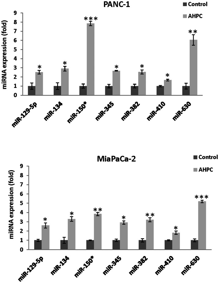
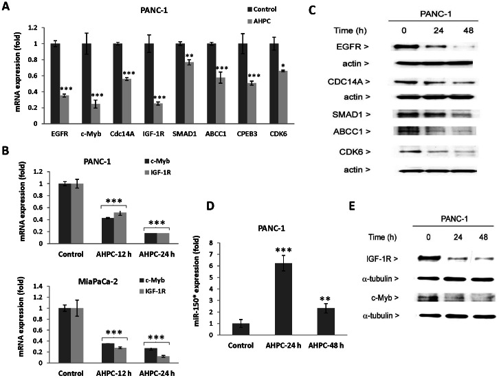
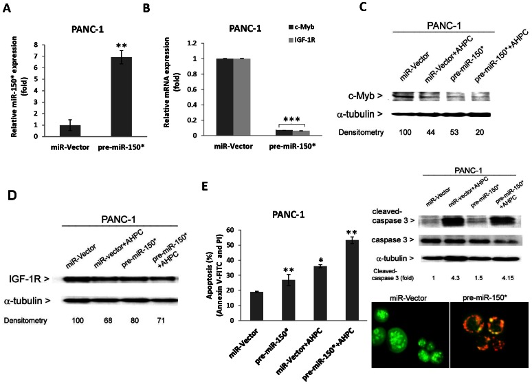
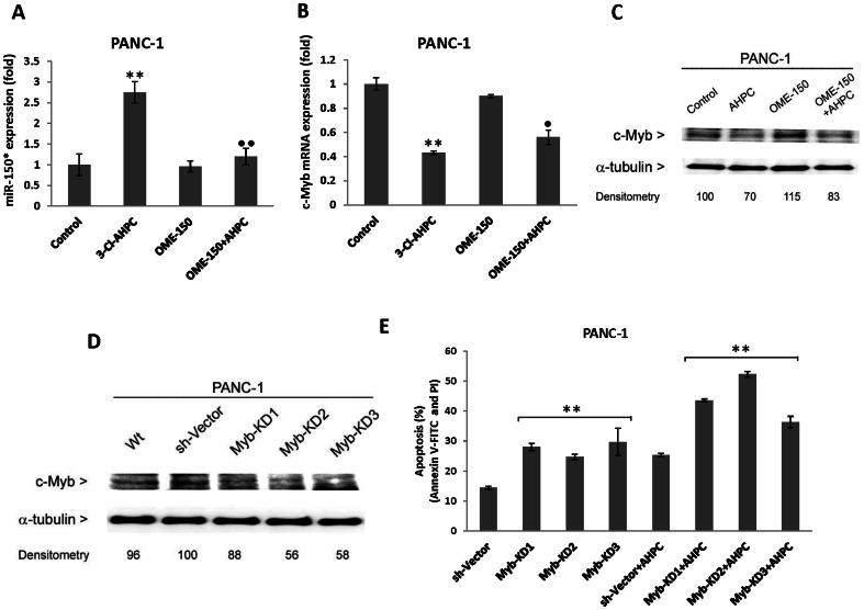
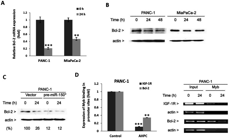
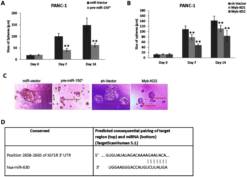
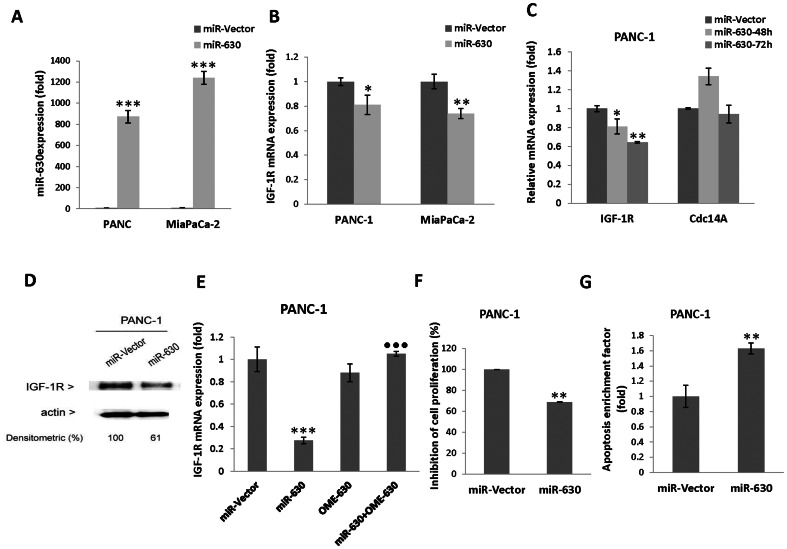
MicroRNAs have been implicated in many critical cellular processes including apoptosis. We have previously found that apoptosis in pancreatic cancer cells was induced by adamantyl retinoid-related (ARR) molecule 3-Cl-AHPC. Here we report that 3-Cl-AHPC-dependent apoptosis involves regulating a number of microRNAs including miR-150* and miR-630. 3-Cl-AHPC stimulated miR-150* expression and caused decreased expression of c-Myb and IGF-1R in the pancreatic cancer cells. 3-Cl-AHPC-mediated reduction of c-Myb resulted in diminished binding of c-Myb with IGF-1R and Bcl-2 promoters, thereby causing repression of their transcription and protein expression. Over-expression of miR-150* also resulted in diminished levels of c-Myb and Bcl-2 proteins. Furthermore, the addition of the miRNA inhibitor 2'-O-methylated miR-150 blocked 3-Cl-AHPC-mediated increase in miR-150* levels and abrogated loss of c-Myb protein. Knockdown of c-Myb in PANC-1 cells resulted in enhanced apoptosis both in the presence or absence of 3-Cl-AHPC confirming the anti-apoptotic property of c-Myb. Overexpression of miR-630 also induced apoptosis in the pancreatic cancer cells and inhibited target protein IGF-1R mRNA and protein expression. Together these results implicate key roles for miR-150* and miR-630 and their targeting of IGF-1R to promote apoptosis in pancreatic cancer cells.


Conflict of interest statement

Competing Interests: The authors have declared that no competing interests exist.

Figures

Figure 1
Figure 1. 3-Cl-AHPC mediated modulation of microRNAs in pancreatic cancer cells.
MicroRNAs were validated by quantitative Real time PCR. Cells were treated with 3-Cl-AHPC for 24 h. Detail methodologies were as described in Materials and Methods. The error bars represent the mean of three separate determinations ± the standard deviation (SD). *, ** and *** (<0.05, <0.01 and <0.001) were significantly different in comparison between control and treated samples using t-Test.
Figure 2
Figure 2. 3-Cl-AHPC mediated expression of microRNA targets in pancreatic cancer cells.
(A and B) Reduced expression of mRNA by SYBR-green RT-PCR; (C and E) decreased proteins levels were demonstrated by Western blots analysis. D). 3-Cl-AHPC exposure increased miR-150* expression and subsequently decreased expression of c-Myb and IGF-1R proteins (E). The error bars represent the mean of three separate determinations ± the standard deviation (SD). *, ** and *** (<0.05, <0.01 and <0.001) were significantly different in comparison between control and treated samples using t-Test.
Figure 3
Figure 3. Overexpression of pre-miR-150* reduced target proteins and induced apoptosis in pancreatic cancer cells.
(A) Increased expression of pre-miR-150* expression vector and pre-miR-150* reduced expression of c-Myb and IGF-1R mRNA and protein in pre-miR-150* stably transfected cells (B–D). Cells were treated with 1 µM 3-Cl-AHPC for 24 h. (E) pre-miR-150* induced apoptosis by itself and enhanced 3-Cl-AHPC mediated apoptosis in PANC-1 cells. Cells were transiently transfected with pre-miR-150* expression vector for 24 h and then exposed to 3-Cl-AHPC for 24 h. Induction of apoptosis was assessed using Annexin V-FITC labeling with propidium iodide (PI) staining in cells and the error bars represent the mean of three separate determinations ± the standard deviation (SD). All treated samples are significantly different from miR-vector. * (<0.05), ** (<0.01 ) and *** (<0.001 ) significantly different in comparison to miR-vector by t-Test. Over-expression of miR-150* induced activation of caspase 3 as indicated by cleaved caspase 3 fragments in pre-miR-150* transiently transfected cells (upper right panel). pre-miR-150* expressed PANC-1 cells induced apoptosis as indicated in acridine orange/ethidium bromide stained cells in lower right panel. Cells were transfected with pre- miR-150* for 48 h and then stained and photographed using Olympus fluorescence microscope digital camera software and DP2-BSW software.
Figure 4
Figure 4. Inhibitor of miR-150* blocked 3-Cl-AHPC-mediated decreased of c-Myb in PANC-1 cells.
(A) Inhibitor 2′-O-methylated miR-150* (OME-150) inhibited 3-Cl-AHPC mediated miR-150* expression in transiently transfected cells. (B and C) miR-150 inhibitor blocked the 3-Cl-AHPC mediated c-Myb degradation. Cells were treated with 1 µM 3-Cl-AHPC for 24 h. (D) Knock down of c-Myb expression in cells. (E) Knock down of c-Myb enhanced the apoptosis in cells. Cells stably transfected with three sh-Myb knockdown expression vectors and scrumble sequence control vector were exposed to 3-Cl-AHPC for 24 h. Induction of apoptosis was assessed using Annexin V-FITC labeling with propidium iodide (PI) staining in cells and the error bars represent the mean of three separate determinations ± the standard deviation (SD). All treated samples and sh-Myb are significantly different from control and sh-vector; • and ••, ** (<0.05 and <0.01 respectively) are significantly different by t-Test and • represented the comparison between 3-Cl-AHPC to OME-150*+3-Cl-AHPC treated samples.
Figure 5
Figure 5. 3-Cl-AHPC mediated decreased c-Myb binding to Bcl-2 and IGF-IR promoters resulted in reduced protein expression.
(A and B) 3-Cl-AHPC decreased Bcl-2 mRNA and protein expression. (C) Over-expression of pre-miR-150* reduced Bcl-2 protein expression. (D) 3-Cl-AHPC decreased c-Myb- binding to IGF-1R and Bcl-2 promoter sites in chromatin immunoprecipitation assay. Cells were exposed to 3-Cl-AHPC for 24 h. The error bars represent the mean of three separate determinations ± the standard deviation (SD). ** and *** (<0.01 and <0.001) were significantly different in comparison between control to 3-Cl-AHPC treated mRNA and Myb binding in promoter sites using t-Test.
Figure 6
Figure 6. Expression of pre-miR-150* and knockdown of c-Myb inhibited CD44+/CD24+ stem-cell like spheres formation in cells.
(A and C) Pre-miR-150* inhibited sphere formation in CD44+/CD24+ cells. (B and C) c-Myb knockdown inhibited sphere formation in CD44+/CD24+ PANC-1 cells. For sphere formation, the CD44+/CD24+ cells were sorted by flow cytometry from stably transfected pre-miR150* and sh-Myb knock down cells, respectively. The sizes of spheres were photographed and measured on a 100 µm scale and magnification 400× using Olympus fluorescence microscope digital camera software and DP2-BSW software. The error bars represent the mean of 15 sphere determinations ± the standard deviation. ** was significantly different in comparison to control spheres. Data were analyzed by ANOVA; Tukey HSD test for multiple comparisons. **P<0.0001 vs control. (D) Scheme showing predicted target conserved site of miR-630 in the 3′UTR of IGF-1R.
Figure 7
Figure 7. Over-expression of pre-miR-630 reduced IGF-1R expression and inhibited cell growth and induced apoptosis in cells.
(A) pre-miR-630 expression in PANC-1 and MiaPaCa-2 cells. (B and C) Increased expression of pre-miR-630 decreased IGF-1R mRNA but not Cdc14A in cells. (D). Decreased IGF-1R protein expression in pre-miR-630 expressed cells. (E) Inhibitor 2′-O-methylated miR-630 (OME-630) blocked the IGF-1R mRNA degradation. Cells were transiently transfected with pre-miR-630 for 48 h (F and G) pre-miR-630 expression inhibited growth and induced apoptosis in PANC-1 cells. Inhibition of cell proliferations was evaluated after 48 h of pre-miR-630 transiently transfected cells by MTT assay and expressed as percent of that inhibited at a time relative to the miR-vector control. The error bars represent the mean of four separate determinations ± the standard deviation (SD). *, ** and *** (<0.05, <0.01 and <0.001) were significantly different in comparison between miR-vector and miR-630; • was highly significant and represented the comparison between miR-630 to miR-630+OME-630 using t-Test. Apoptosis of miR-630 cells was evaluated after 48 h of transfected cells. The cell death was measured by cytoplasmic histone-associated-DNA-fragments by ELISA (enrichment factor = OD of miR-630 expressed lysate/OD of miR-vector, OD at 405 nm-490 nm).

Similar articles

Cited by

References

    1. Lee YS, Dutta A (2009) MicroRNAs in cancer. Annu Rev Pathol 4: 199–227. - PMC - PubMed
    1. Nanna-Sinkam SP, Croce CM (2011) Non-coding RNAs in cancer initiation and progression and as novel biomarkers. Mol Oncol 5: 483–491. - PMC - PubMed
    1. Bhayani MK, Calin GA, Lai SY (2012) Functional relevance of miRNA sequences in human disease. Mutat Res 731: 14–19. - PMC - PubMed
    1. Mott JL (2009) Micro RNAs involved in tumor suppressor and oncogene pathways: Implication for hepatobiliary neoplasia. Hepatology 50: 630–637. - PMC - PubMed
    1. Bartel DP (2009) MicroRNAs. Target recognition and regulatory functions. Cell 136: 215–233. - PMC - PubMed

Publication types

MeSH terms