. 2013 May 13;8(5):e62659.
doi: 10.1371/journal.pone.0062659. Print 2013.

Basolateral BMP signaling in polarized epithelial cells

Affiliations

Basolateral BMP signaling in polarized epithelial cells

Masao Saitoh et al. PLoS One. .

Abstract

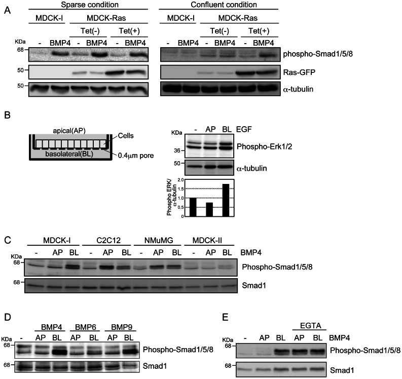
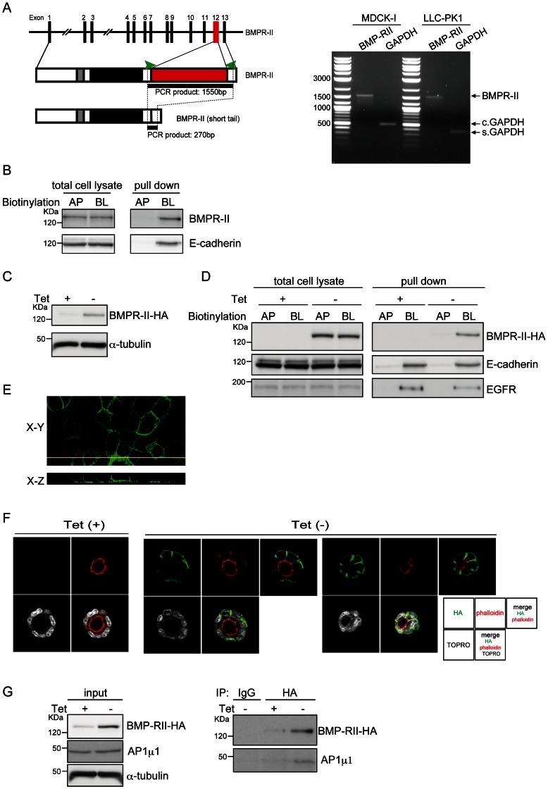
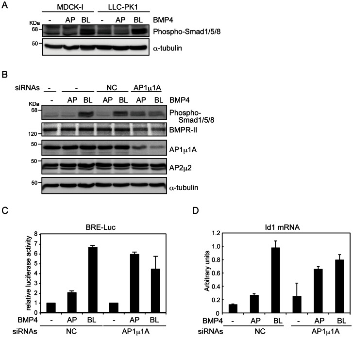
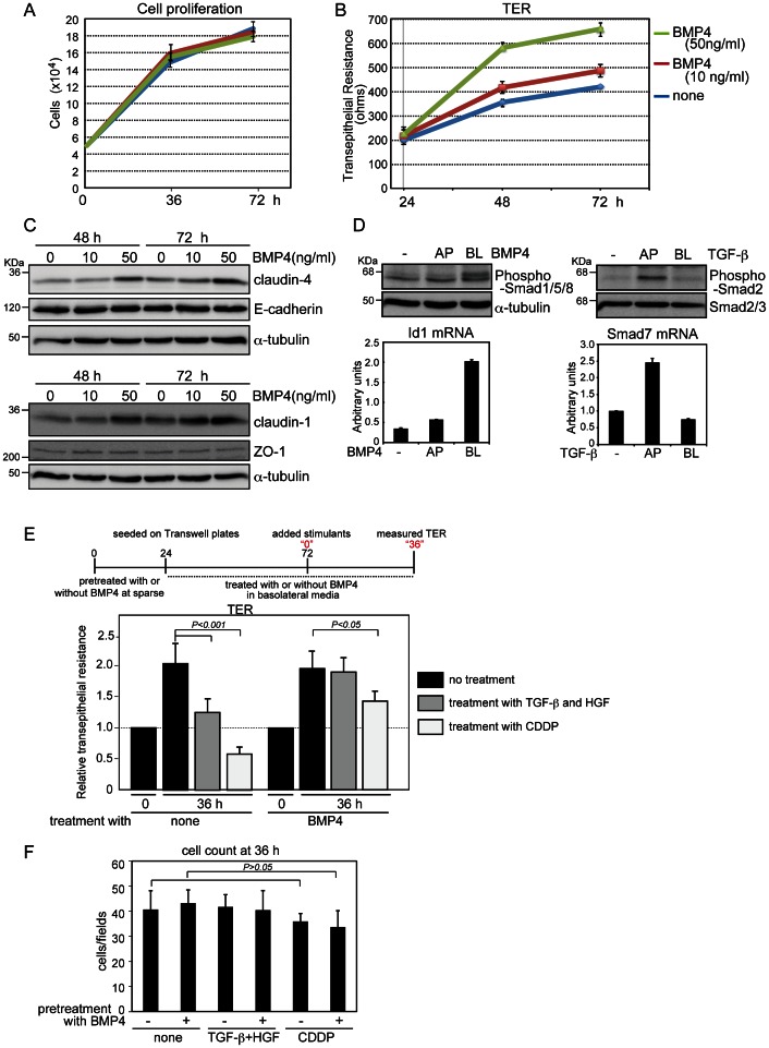
Bone morphogenetic proteins (BMPs) regulate various biological processes, mostly mediated by cells of mesenchymal origin. However, the roles of BMPs in epithelial cells are poorly understood. Here, we demonstrate that, in polarized epithelial cells, BMP signals are transmitted from BMP receptor complexes exclusively localized at the basolateral surface of the cell membrane. In addition, basolateral stimulation with BMP increased expression of components of tight junctions and enhanced the transepithelial resistance (TER), counteracting reduction of TER by treatment with TGF-β or an anti-tumor drug. We conclude that BMPs maintain epithelial polarity via intracellular signaling from basolaterally localized BMP receptors.


Conflict of interest statement

Competing Interests: The authors have declared that no competing interests exist.

Figures

Figure 1
Figure 1. BMP signaling from the basolateral side in polarized epithelial cells.
(A) Phosphorylation of Smad1/5/8 induced by BMP4 was examined by immunoblot analyses. MDCK-I and MDCK-Ras cells were seeded at the density of 2.0×10⁵ (sparse) or 1.0×10⁶ (confluent) cells in 6-well plates. Twenty four hours later, the cells were treated with 20 ng/ml BMP4 for 45 min. (B) MDCK-I cells were grown to confluence on Transwell plates (left) and treated with 10 ng/ml of EGF from the apical (AP) or basolateral (BL) sides for 15 min. Phosphorylation of Erk1/2 induced by EGF was examined by immunoblot analyses. The ratio of phospho-Erk1/2 to α-tubulin was validated by densitometric analysis and shown at the bottom. (C, D, and E) MDCK-I, C2C12, NMuMG, and MDCK-II cells were grown to confluence on Transwell plates and treated with BMPs from the apical (AP) or basolateral (BL) sides for 45 min. Phosphorylation of Smad1/5/8 induced by BMP4 (20 ng/ml), BMP6 (20 ng/ml), and BMP9 (2 ng/ml) was examined by immunoblot analyses. EGTA (1 mM) was added 3 h before treatment with BMP4 (E).
Figure 2
Figure 2. Basolateral localization of BMPR-II.
(A) A schematic illustration of the BMPR-II gene and protein, containing the transmembrane (grey) and kinase (black) domains and the region encoded by alternative exon 12 (red), is shown on the left. The primers used for PCR are shown as green arrowheads. Expression of alternative-splicing variants of BMPR-II in MDCK-I and LLC-PK1 cells was examined by RT-PCR. GAPDH was used as internal control for Canis lupus familiaris MDCK-I cells (c. GAPDH) and Sus scrofa LLC-PK1 cells (s. GAPDH). (B) After MDCK-I cells cultured on Transwells in a confluent condition were biotinylated from apical (AP) or basolateral (BL) sides, equal amounts of total proteins from each surface were subjected to SDS-PAGE (total cell lysate), or incubated with Streptavidin Sepharose 4B to isolate biotinylated proteins (pulldown). E-cadherin, a representative basolateral protein, was used as a control. (C) After withdrawal of tetracycline (Tet) from the culture media, MDCK-BR2 cells were lysed and subjected to immunoblot analysis with an antibody against HA. (D) MDCK-BR2 cells were seeded on Transwell plates to reach confluence in the presence or absence of tetracycline (Tet), and biotinylated from the apical (AP) or basolateral (BL) sides. Equal quantities of proteins were subjected to SDS-PAGE (total cell lysate), or incubated with Streptavidin Sepharose 4B to isolate biotinylated proteins, followed by SDS-PAGE (pulldown). E-cadherin and EGFR were used as representative basolateral proteins. (E) MDCK-BR2 cells were stained with an anti-HA antibody (green). XY (horizontal) and XZ (vertical) sections are shown in the top and bottom panels, respectively. (F) MDCK-BR2 cells cultured in Matrigel in the presence or absence of tetracycline (Tet) were stained with an anti-HA antibody (green), rhodamine-phalloidin (red), and TOPRO (white), followed by fluorescence imaging using a confocal laser scanning microscope. Micrographs of the two independent colonies were taken in the absence of Tet. (G) After MDCK-BR2 cells were cultured in the presence or absence of tetracycline (Tet), cell lysates were assayed for protein concentration and then subjected to immunoprecipitation. Co-purified AP1 µ1 was detected by immunoblotting with an anti-AP1 µ1 antibody. The expression levels of BMPR-II-HA, AP1 µ1, and α-tubulin in the same lysates were verified by immunoblotting.
Figure 3
Figure 3. Roles of AP1 µ1A in basolateral trafficking of BMPR-II.
(A and B) MDCK-I cells and LLC-PK1 cells transfected with either control (NC) or AP1 µ1A siRNAs were grown to confluence on Transwell plates, and then treated with 20 ng/ml BMP4 from the apical (AP) or basolateral (BL) sides for 45 min. Phosphorylation of Smad1/5/8 induced by BMP4 was examined by immunoblot analyses using the indicated antibodies. (C and D) LLC-PK1 cells and LLC-PK1 cells transfected with BMP-responsive element (BRE)-reporter construct in combination with either control siRNA (NC) or AP1 µ1A siRNA were seeded on Transwell plates and grown to confluence. Cells were treated with BMP4 (20 ng/ml) from the apical (AP) or basolateral (BL) sides for 2 h for luciferase assays, normalized to activity of sea pansy luciferase encoded by pRL-TK-Renilla (C), and for 1 h for quantitative RT-PCR analysis, normalized to the amounts of GAPDH mRNA (D). Each value represents the mean ± S.D. of triplicate determinations from a representative experiment. Similar results were obtained in two independent experiments (C and D).
Figure 4
Figure 4. Increase of transepithelial resistance (TER) by BMP4 treatment.
(A, B, and C) MDCK-I cells pretreated with BMP4 for 24 h were seeded on Transwell plates in basolateral media containing BMP4, and incubated until 72 h (i.e., for an additional 48 h). Cell counting (A), TER measurement (B), and immunoblot analyses (C) were performed at the indicated time points. (D) MDCK-I cells were grown to confluence on Transwell plates and treated for 45 min with 20 ng/ml BMP4 (left) and 1 ng/ml TGF-β (right) from the apical (AP) or basolateral (BL) sides. Phosphorylation of Smads and expression of representative target genes (Id1 for BMP4 and Smad7 for TGF-β) were examined by immunoblot and quantitative RT-PCR analyses, respectively. Each value represents the mean ± S.D. of triplicate determinations from a representative experiment. Similar results were obtained in two independent experiments (A, B and D). (E and F) MDCK-I cells pretreated with BMP4 under sparse conditions for 24 h were seeded in triplicate on Transwell plates in basolateral media containing 50 ng/ml BMP4 for 48 h, and TER was measured (indicated as “0”). Then the cells were treated for 36 h either with both 1 ng/ml TGF-β from the apical side and 10 ng/ml HGF from the basolateral side or with 25 µM CDDP from the basolateral side. After TER of all three Transwell plates had been measured at four points for each well (E), the cells from two plates were used for cell counting (F) and the other was used for E-cadherin staining (Fig. S3). Each value represents the mean±S.D. of duplicate determinations from a representative experiment. Similar results were obtained in three independent experiments.

Similar articles

Cited by

References

    1. Keita AV, Soderholm JD (2012) Barrier dysfunction and bacterial uptake in the follicle-associated epithelium of ileal Crohn’s disease. Ann. N. Y. Acad. Sci. 1258: 125–134. - PubMed
    1. Zeisberg M, Bonner G, Maeshima Y, Colorado P, Muller GA, et al. (2001) Renal fibrosis: collagen composition and assembly regulates epithelial-mesenchymal transdifferentiation. Am. J. Pathol. 159: 1313–1321. - PMC - PubMed
    1. Tsukita S, Furuse M, Itoh M (2001) Multifunctional strands in tight junctions. Nat. Rev. Mol. Cell Biol. 2: 285–293. - PubMed
    1. Rodriguez-Boulan E, Kreitzer G, Musch A (2005) Organization of vesicular trafficking in epithelia. Nat. Rev. Mol. Cell Biol. 6: 233–247. - PubMed
    1. Folsch H (2005) The building blocks for basolateral vesicles in polarized epithelial cells. Trends Cell Biol. 15: 222–228. - PubMed

Publication types

MeSH terms

LinkOut - more resources