

Color Doppler imaging analysis of retrobulbar blood flow velocities in primary open-angle glaucomatous eyes: a meta-analysis
- PMID: 23675419
- PMCID: PMC3652862
- DOI: 10.1371/journal.pone.0062723
Color Doppler imaging analysis of retrobulbar blood flow velocities in primary open-angle glaucomatous eyes: a meta-analysis
Abstract
Background: To analyze the diagnostic value of color Doppler imaging (CDI) of blood flow in the retrobulbar vessels of eyes with primary open-angle glaucoma (POAG).
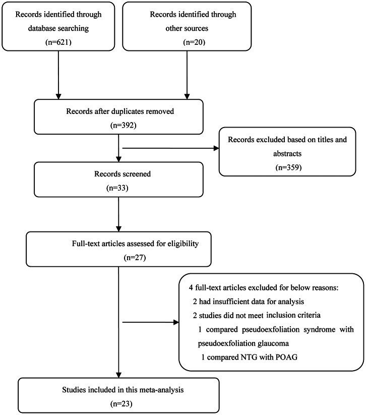
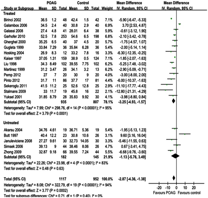
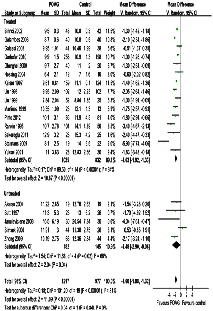
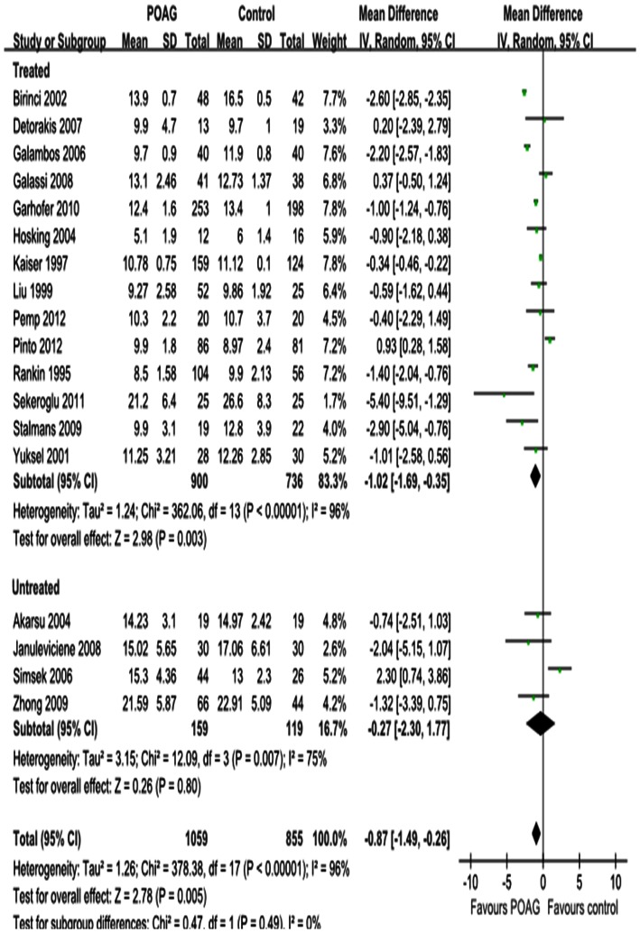
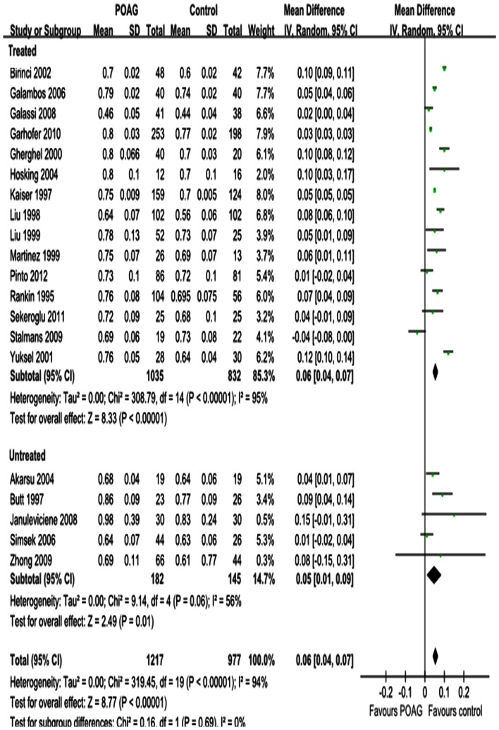
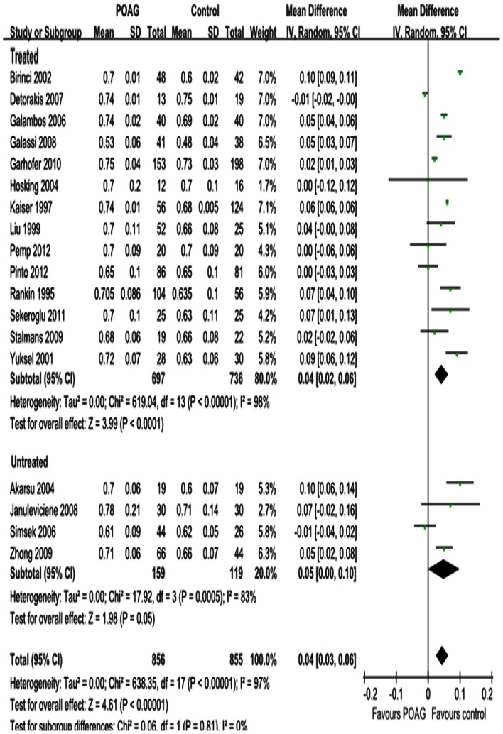
Methods: Pertinent publications were retrieved from the Cochrane Central Register of Controlled Trials, PubMed and the ISI Web of Knowledge up to October 2012. Changes in peak systolic velocity (PSV), end diastolic velocity (EDV) and resistive index (RI) of the ophthalmic artery (OA), central retinal artery (CRA) and short posterior ciliary artery (SPCA) of POAG eyes and normal controls were evaluated by CDI. Subgroup analyses were conducted according to whether patients received IOP-lowering drugs treatment and were defined as treated and untreated.
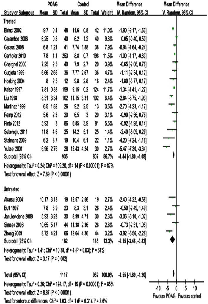
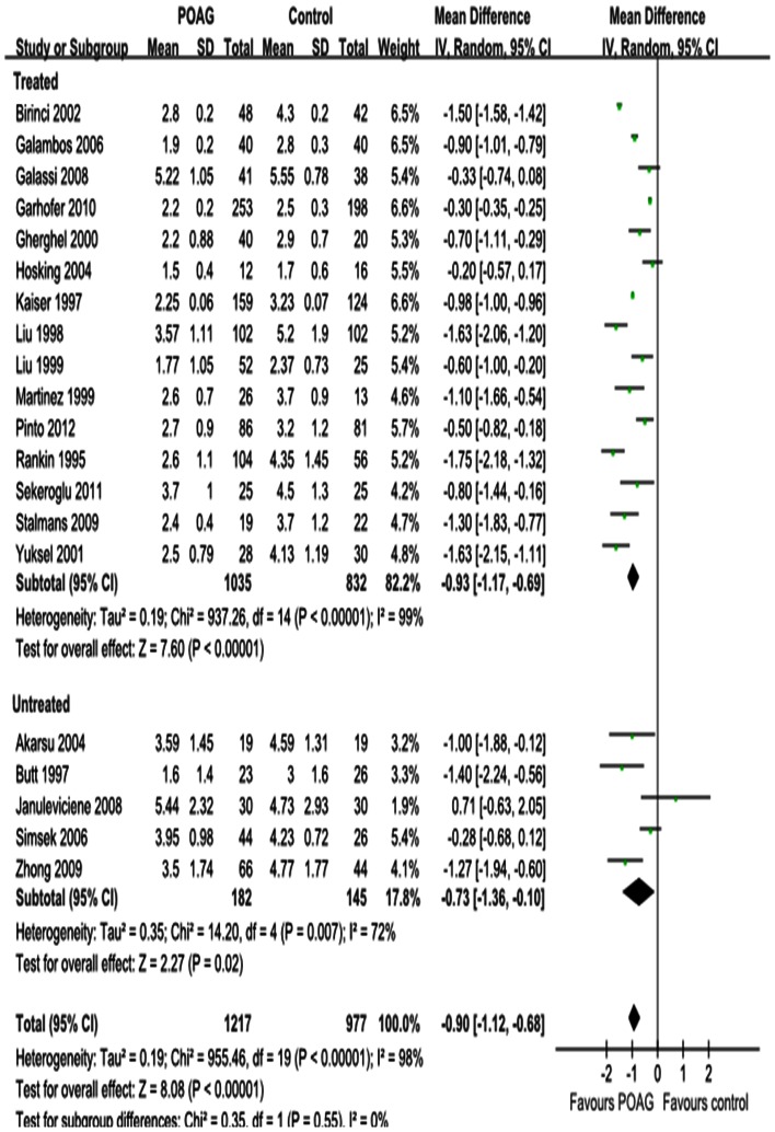
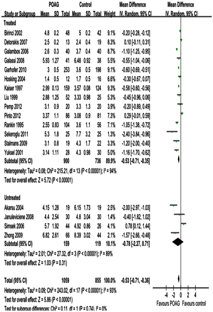
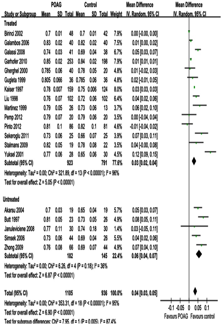
Results: PSV and EDV were statistically significantly reduced in the OA of POAG eyes (P = 0.0002; P<0.00001; respectively), with significant heterogeneity (P(heterogeneity)<0.00001, I² = 94%; P(heterogeneity)<0.00001, I² = 85%; respectively). Similar results were demonstrated for the CRA (P<0.00001; respectively) and SPCA (P = 0.005; P<0.00001; respectively), with significant heterogeneities for both the CRA (P(heterogeneity)<0.00001, I² = 81%; P(heterogeneity)<0.00001, I² = 98%; respectively) and the SPCA (P(heterogeneity)<0.00001, I² = 96%; P(heterogeneity)<0.00001, I² = 93%; respectively). Significant increases in RI were found in all retrobulbar vessels (P<0.00001; respectively), with significant heterogeneities (P(heterogeneity)<0.00001, I² = 95%; P(heterogeneity)<0.00001, I² = 94%; P(heterogeneity)<0.00001, I² = 97%; respectively).
Conclusions: This meta-analysis suggests that CDI is a potential diagnostic tool for POAG.
Conflict of interest statement
Figures
Similar articles
-
Assessment of orbital hemodynamic changes in primary open angle glaucoma by color Doppler imaging.J Fr Ophtalmol. 2023 Nov;46(9):1061-1068. doi: 10.1016/j.jfo.2023.03.018. Epub 2023 Aug 23. J Fr Ophtalmol. 2023. PMID: 37625998
-
Color Doppler imaging and pattern visual evoked potential in normal tension glaucoma and hypertension glaucoma.Doc Ophthalmol. 2009 Dec;119(3):171-80. doi: 10.1007/s10633-009-9192-7. Epub 2009 Sep 12. Doc Ophthalmol. 2009. PMID: 19756801
-
Correlation of retrobulbar perfusion deficits with glaucomatous visual field defects.Graefes Arch Clin Exp Ophthalmol. 2024 Sep;262(9):2961-2970. doi: 10.1007/s00417-024-06464-3. Epub 2024 Apr 8. Graefes Arch Clin Exp Ophthalmol. 2024. PMID: 38587654
-
Color Doppler imaging of the retrobulbar circulation in glaucoma.Surv Ophthalmol. 1999 Jun;43 Suppl 1:S176-82. doi: 10.1016/s0039-6257(99)00043-0. Surv Ophthalmol. 1999. PMID: 10416761 Review.
-
Feasibility of creating a normative database of colour Doppler imaging parameters in glaucomatous eyes and controls.Br J Ophthalmol. 2011 Sep;95(9):1193-8. doi: 10.1136/bjo.2010.188219. Epub 2010 Nov 24. Br J Ophthalmol. 2011. PMID: 21106991 Review.
Cited by
-
Glaucoma Surgery and Ocular Blood Flow in Colour Doppler Imaging: Is There a Link?Clin Ophthalmol. 2024 Jan 6;18:49-60. doi: 10.2147/OPTH.S441805. eCollection 2024. Clin Ophthalmol. 2024. PMID: 38205265 Free PMC article. Review.
-
Ocular blood flow and choroidal thickness in ocular hypertension.Int Ophthalmol. 2022 May;42(5):1357-1368. doi: 10.1007/s10792-021-02123-2. Epub 2021 Nov 25. Int Ophthalmol. 2022. PMID: 34822054
-
Classification of optic disc shape in glaucoma using machine learning based on quantified ocular parameters.PLoS One. 2017 Dec 19;12(12):e0190012. doi: 10.1371/journal.pone.0190012. eCollection 2017. PLoS One. 2017. PMID: 29261773 Free PMC article.
-
Does steep Trendelenburg positioning effect the ocular hemodynamics and intraocular pressure in patients undergoing robotic cystectomy and robotic prostatectomy?Int Urol Nephrol. 2017 Jan;49(1):55-60. doi: 10.1007/s11255-016-1449-y. Epub 2016 Nov 1. Int Urol Nephrol. 2017. PMID: 27804081
-
The diagnostic use of choroidal thickness analysis and its correlation with visual field indices in glaucoma using spectral domain optical coherence tomography.PLoS One. 2017 Dec 13;12(12):e0189376. doi: 10.1371/journal.pone.0189376. eCollection 2017. PLoS One. 2017. PMID: 29236748 Free PMC article.
References
-
- Van Buskirk EM, Cioffi GA (1992) Glaucomatous optic neuropathy. Am J Ophthalmol 113: 447–452. - PubMed
-
- Resch H, Garhofer G, Fuchsjager-Mayrl G, Hommer A, Schmetterer L (2009) Endothelial dysfunction in glaucoma. Acta Ophthalmol 87: 4–12. - PubMed
-
- Flammer J, Orgul S, Costa VP, Orzalesi N, Krieglstein GK, et al. (2002) The impact of ocular blood flow in glaucoma. Prog Retin Eye Res 21: 359–393. - PubMed
-
- Costa VP, Harris A, Stefansson E, Flammer J, Krieglstein GK, et al. (2003) The effects of antiglaucoma and systemic medications on ocular blood flow. Prog Retin Eye Res 22: 769–805. - PubMed
Publication types
MeSH terms
LinkOut - more resources
Full Text Sources
Other Literature Sources
Medical
