

The impact of genetic susceptibility to systemic lupus erythematosus on placental malaria in mice
- PMID: 23675429
- PMCID: PMC3651086
- DOI: 10.1371/journal.pone.0062820
The impact of genetic susceptibility to systemic lupus erythematosus on placental malaria in mice
Abstract
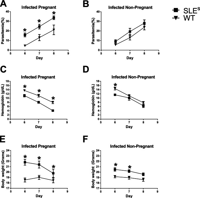
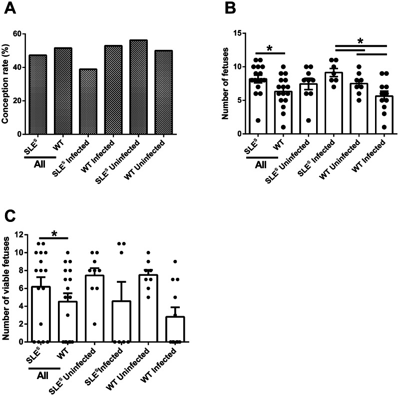
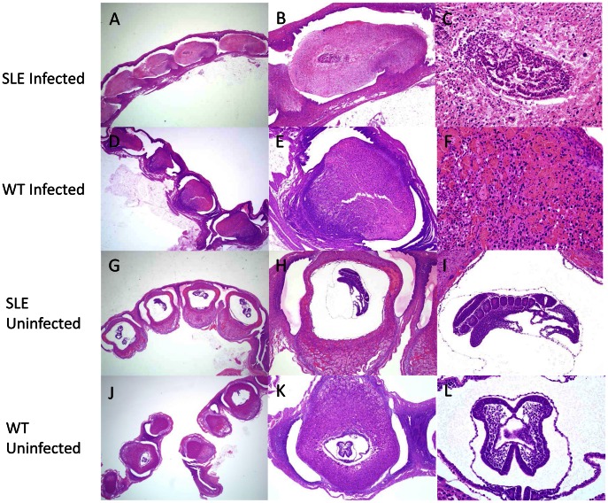
Severe malaria, including cerebral malaria (CM) and placental malaria (PM), have been recognized to have many of the features of uncontrolled inflammation. We recently showed that in mice genetic susceptibility to the lethal inflammatory autoimmune disease, systemic lupus erythematosus (SLE), conferred resistance to CM. Protection appeared to be mediated by immune mechanisms that allowed SLE-prone mice, prior to the onset of overt SLE symptoms, to better control their inflammatory response to Plasmodium infection. Here we extend these findings to ask does SLE susceptibility have 1) a cost to reproductive fitness and/or 2) an effect on PM in mice? The rates of conception for WT and SLE susceptible (SLE(s)) mice were similar as were the number and viability of fetuses in pregnant WT and SLE(s) mice indicating that SLE susceptibility does not have a reproductive cost. We found that Plasmodium chabaudi AS (Pc) infection disrupted early stages of pregnancy before the placenta was completely formed resulting in massive decidual necrosis 8 days after conception. Pc-infected pregnant SLE(s) mice had significantly more fetuses (∼1.8 fold) but SLE did not significantly affect fetal viability in infected animals. This was despite the fact that Pc-infected pregnant SLE(s) mice had more severe symptoms of malaria as compared to Pc-infected pregnant WT mice. Thus, although SLE susceptibility was not protective in PM in mice it also did not have a negative impact on reproductive fitness.
Conflict of interest statement
Figures
Similar articles
-
Murine malaria infection induces fetal loss associated with accumulation of Plasmodium chabaudi AS-infected erythrocytes in the placenta.Infect Immun. 2006 May;74(5):2839-48. doi: 10.1128/IAI.74.5.2839-2848.2006. Infect Immun. 2006. PMID: 16622222 Free PMC article.
-
Composition of the gut microbiota transcends genetic determinants of malaria infection severity and influences pregnancy outcome.EBioMedicine. 2019 Jun;44:639-655. doi: 10.1016/j.ebiom.2019.05.052. Epub 2019 May 31. EBioMedicine. 2019. PMID: 31160271 Free PMC article.
-
Systemic lupus erythematosus-associated defects in the inhibitory receptor FcgammaRIIb reduce susceptibility to malaria.Proc Natl Acad Sci U S A. 2007 Apr 24;104(17):7169-74. doi: 10.1073/pnas.0608889104. Epub 2007 Apr 13. Proc Natl Acad Sci U S A. 2007. PMID: 17435165 Free PMC article.
-
Malaria in pregnancy: linking immunity and pathogenesis to prevention.Am J Trop Med Hyg. 2007 Dec;77(6 Suppl):14-22. Am J Trop Med Hyg. 2007. PMID: 18165470 Review.
-
Genome screening for susceptibility loci in systemic lupus erythematosus.Am J Pharmacogenomics. 2002;2(1):1-12. doi: 10.2165/00129785-200202010-00001. Am J Pharmacogenomics. 2002. PMID: 12083950 Review.
Cited by
-
Tempol, an intracellular antioxidant, inhibits tissue factor expression, attenuates dendritic cell function, and is partially protective in a murine model of cerebral malaria.PLoS One. 2014 Feb 28;9(2):e87140. doi: 10.1371/journal.pone.0087140. eCollection 2014. PLoS One. 2014. PMID: 24586264 Free PMC article.
References
-
- Umbers AJ, Aitken EH, Rogerson SJ (2011) Malaria in pregnancy: small babies, big problem. Trends Parasitol 27: 168–175. - PubMed
-
- Brabin BJ, Romagosa C, Abdelgalil S, Menendez C, Verhoeff FH, et al. (2004) The sick placenta-the role of malaria. Placenta 25: 359–378. - PubMed
-
- Molokhia M, McKeigue P (2006) Systemic lupus erythematosus: genes versus environment in high risk populations. Lupus 15: 827–832. - PubMed
-
- Bae SC, Fraser P, Liang MH (1998) The epidemiology of systemic lupus erythematosus in populations of African ancestry: a critical review of the “prevalence gradient hypothesis”. Arthritis Rheum 41: 2091–2099. - PubMed
Publication types
MeSH terms
Grants and funding
LinkOut - more resources
Full Text Sources
Other Literature Sources
Medical
Molecular Biology Databases
Research Materials
