. 2013 May 13;8(5):e62926.
doi: 10.1371/journal.pone.0062926. Print 2013.

A soft coral-derived compound, 11-epi-sinulariolide acetate suppresses inflammatory response and bone destruction in adjuvant-induced arthritis

Affiliations

A soft coral-derived compound, 11-epi-sinulariolide acetate suppresses inflammatory response and bone destruction in adjuvant-induced arthritis

Yen-You Lin et al. PLoS One. .

Abstract

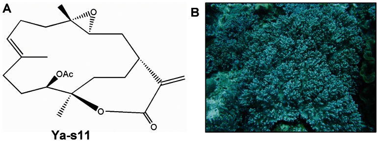
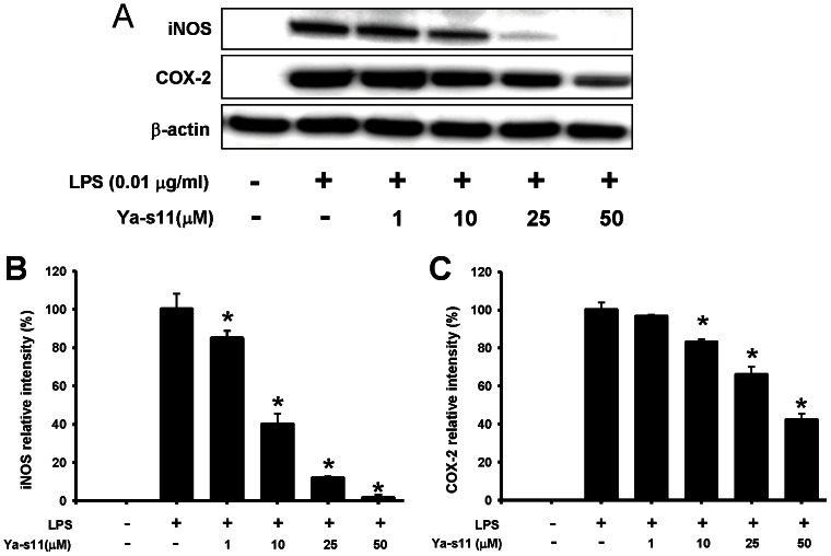
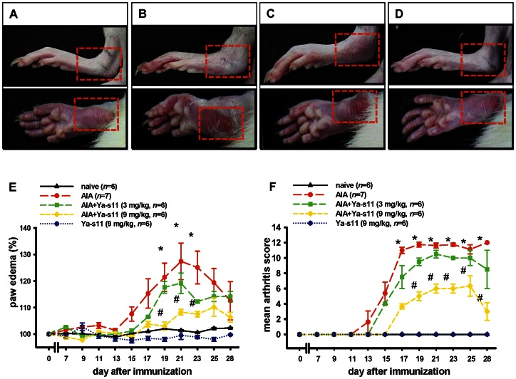
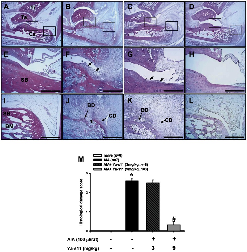
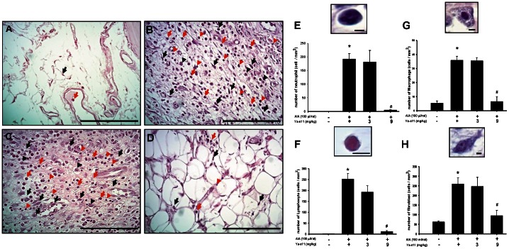
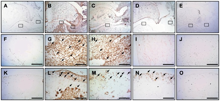
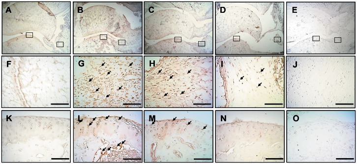
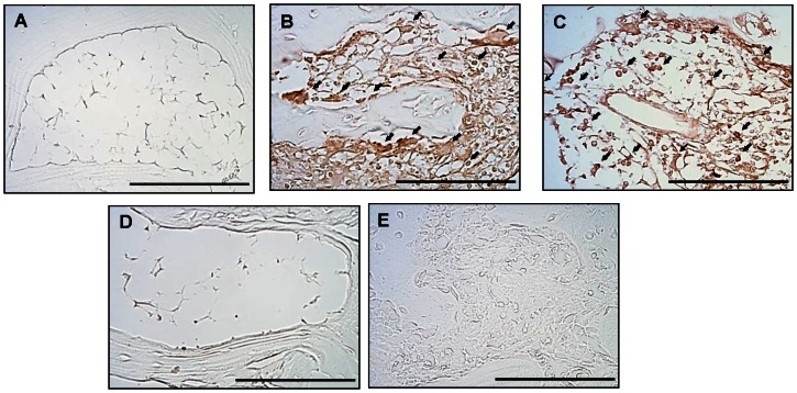
In recent years, a significant number of metabolites with potent anti-inflammatory properties have been discovered from marine organisms, and several of these compounds are now under clinical trials. In the present study, we isolated 11-epi-sinulariolide acetate (Ya-s11), a cembrane-type compound with anti-inflammatory effects, from the Formosa soft coral Sinularia querciformis. Preliminary screening revealed that Ya-s11 significantly inhibited the expression of the proinflammatory proteins induced nitric oxide synthase and cyclooxygenase-2 in lipopolysaccharide-stimulated murine macrophages. We also examined the therapeutic effects of Ya-s11 on adjuvant-induced arthritis (AIA) in female Lewis rats, which demonstrate features similar to human rheumatoid arthritis (RA). Animal experiments revealed that Ya-s11 (subcutaneously 9 mg/kg once every 2 days from day 7 to day 28 postimmunization) significantly inhibited AIA characteristics. Moreover, Ya-s11 also attenuated protein expression of cathepsin K, matrix metalloproteinases-9 (MMP-9), tartrate-resistant acid phosphatase (TRAP), and tumor necrosis factor-α (TNF-α) in ankle tissues of AIA-rats. Based on its attenuation of the expression of proinflammatory proteins and disease progression in AIA rats, the marine-derived compound Ya-s11 may serve as a useful therapeutic agent for the treatment of RA.


Conflict of interest statement

Competing Interests: The authors have declared that no competing interests exist.

Figures

Figure 1
Figure 1. Chemical structure and source of 11-epi-sinulariolide acetate (Ya-s11).
(A) The chemical structure of 11-epi-sinulariolide acetate. Molecular formula, C22H32O5, Molecular weight, 376. (B) The soft coral sample, Sinularia querciformis, was collected from Dongsha Island.
Figure 2
Figure 2. Effect of Ya-s11 on pro-inflammatory iNOS and COX-2 protein expression in LPS-stimulated macrophage cells.
(A) immunoreactive bands corresponding to iNOS, COX-2, and β-actin protein from RAW 264.7 cells; (B) relative density of iNOS immunoreactive bands; (C) relative density of COX-2 immunoreactive bands. The relative intensity of the LPS group was set to 100%. Band intensities were quantified by densitometry and are indicated as the percent change relative to that of the LPS group. Western blotting with β-actin was performed to verify that equivalent amounts of protein were loaded in each lane. Ya-s11 significantly inhibited LPS-induced iNOS and COX-2 protein expression in murine Raw 264.7 macrophage cells. The experiment was repeated 4 times. *P<0.05, significantly different from the LPS-induced group.
Figure 3
Figure 3. Effect of Ya-s11 on AIA rats.
Typical representative macroscopic photographs of ankle and paw from the (A) naïve, (B) AIA, (C) AIA+Ya−s11 (3 mg/kg), and (D) AIA+Ya−s11 (9 mg/kg) groups. The AIA and AIA+Ya−s11 (3 mg/kg) groups displayed significant the edema on ankle joints and erythema on hindpaws (red square) compared to the naïve group (C). The AIA+Ya−s11 (9 mg/kg) group demonstrated apparent reduction of AIA-induced edema and erythema (D). Quantitative analysis of the effect of Ya-s11 at doses of 3 or 9 mg/kg on AIA-induced paw edema (E). Baseline values for the paw volume of each rat were set to 100%, and changes in edema level were calculated as a percentage increase from the control (pre-drug) volume. Ya-s11 demonstrated dose-dependent inhibition of AIA-induced paw edema in rats. Clinical evaluation of the effect of Ya-s11 on AIA-induced clinical signs in rats (F). Ya-s11 demonstrated a dose-dependent effect on AIA-induced clinical signs. Ya-s11 (3 or 9 mg/kg) was subcutaneously injected every 2 days between day 7 and day 28. Values reflect the mean ± SEM for each group. *P<0.05 compared with the naïve group. # P<0.05 compared with the AIA group.
Figure 4
Figure 4. Histopathological assessments of the effect of Ya-s11 on the AIA rat ankle joint.
Representative sections of ankle joint from the (A, E, I) naïve, (B, F, J) AIA, (C, G, K) AIA+Ya-s11 (3 mg/kg), and (D, H, L) AIA+Ya-s11 (9 mg/kg) groups stained with H&E. Normal joint structure showing calcaneus-talus articulation with the distal tibia and normal synovial tissue was observed in the naïve group (A). Marked joint destruction with bone damage, synovial tissue hyperplasia, and increased relative size of the marrow cavity of the subchondral bone marrow was observed in the AIA group (B). A higher-magnification view of AIA group shows pannus formation (arrow) and bone erosion over the rim of articular bone (F), and synovial tissue cells infiltration into subchondral bone marrow through the erosive orifice (arrowhead) into the calcaneus (J). The AIA+Ya-s11 (3 mg/kg) group demonstrated marked joint destruction with bone damage and synovial tissue hyperplasia (C). A higher-magnification view of AIA+Ya-s11 (3 mg/kg) group shows less severe pannus formation (arrow) and cartilage and bone destruction and resorption on the calcaneus compared with the AIA group (G). In the AIA+Ya-s11 (9 mg/kg) group, no morphological change in the ankle joint was apparent (H, L). The representative histopathological scores (M) of each group were analyzed to assess the degree of morphological changes, and the AIA+Ya-s11 (9 mg/kg) group demonstrated a significant decrease in the degree of arthritis. SB, subchondral bone marrow; Ca, calcaneus; Ta, talus; Ti, tibia; CD, cartilage destruction; BD, bone destruction; E–L, scale bar = 500 µm. *P<0.05 compared with the naïve group. # P<0.05 compared with the AIA group.
Figure 5
Figure 5. Effect of Ya-s11 on cell infiltration to the synovial tissue in AIA rats.
Representative photographs of H&E-stained synovial tissue from the (A) naïve, (B) AIA, (C) AIA+Ya-s11 (3 mg/kg), and (D) AIA+Ya-s11 (9 mg/kg) groups. The synovial tissue of the naïve group demonstrated synovial fibroblasts with few immune cells (arrow) (A). Upregulation of neutrophils (red arrow), lymphocytes (red head arrow), macrophages (black head arrow), and synovial fibroblasts (black arrow) was observed in synovial tissue from the AIA group (B) and AIA+Ya-s11 (3 mg/kg) group (C). Ya-s11 (9 mg/kg) appeared to attenuate AIA-induced upregulation of immune cells. The numbers of neutrophils (E), lymphocytes (F), macrophages (G), and synovial fibroblasts (H) were analyzed in synovial tissue from each group. Higher-magnification views of synovial tissue from the AIA group shows histopathological features of infiltrating cells (E-H). Cell numbers significantly increased between the naïve group and AIA group and were significantly decreased in the AIA+Ya-s11 (9 mg/kg) group compared with the AIA group. * P<0.05 compared with the naïve group. # P<0.05 compared with the AIA group. A–D, scale bar = 100 µm; E–H, scale bar = 5 µm.
Figure 6
Figure 6. Effect of Ya-s11 on cathepsin K protein expression in the ankle of AIA rats.
Cathepsin K protein immunoreactivity is shown in red-brown (arrow) in ankle joint sections from the (A, F, K) naïve, (B, G, L) AIA, (C, H, M) AIA+Ya-s11 (3 mg/kg), and (D, I, N) AIA+Ya-s11 (9 mg/kg) groups. (F)–(J) show cathepsin K immunoreactivity in the synovial tissue of ankle joints outlined in boxes in (A)–(E), respectively. (K)–(O) show cathepsin K immunoreactivity in the articular cartilage outlines in boxes in (A)–(E), respectively. The immunostaining results indicate the upregulation of cathepsin K protein expression in the ankle joint (synovial tissue and cartilage) in AIA rats (B, G, L) and the inhibition of cathepsin K protein expression in synovial tissue and cartilage by treatment with 9 mg/kg but not 3 mg/kg Ya-s11. (E, J, O) A sample from the AIA group incubated without primary antibody for cathepsin K showed no specific staining. Scale bar = 100 µm.
Figure 7
Figure 7. Effect of Ya-s11 on MMP-9 protein expression in the ankle joint of AIA rats.
MMP-9 protein immunoreactivity is shown in red-brown (arrow) in ankle joint sections from the (A, F, K) naïve, (B, G, L) AIA, (C, H, M) AIA+Ya−s11 (3 mg/kg), and (D, I, N) AIA+Ya−s11 (9 mg/kg) groups. (F)–(J) show MMP-9 immunoreactivity in the synovial tissue of ankle joints outlined with boxes in (A)–(E), respectively. (K)–(O) show MMP-9 immunoreactivity in the articular cartilage outlined with boxes in (A)–(E), respectively. The immunostaining results indicate the upregulation of MMP-9 protein expression in the ankle joint (synovial tissue and cartilage) in AIA rat (B, G, L) and inhibition of the AIA-induced upregulation of MMP-9 protein expression in synovial tissue and cartilage by treatment with 9 mg/kg but not 3 mg/kg Ya-s11. (E, J, O) A sample from the AIA group incubated without primary antibody for MMP-9 showed no specific staining. Scale bar = 100 µm.
Figure 8
Figure 8. Effect of Ya-s11 on TRAP protein expression in subchondral bone marrow of AIA rats.
TRAP protein immunoreactivity is shown in red-brown (arrow) in the bone marrow of ankle joint sections from the (A) naïve, (B) AIA, (C) AIA+Ya−s11 (3 mg/kg), and (D) AIA+Ya−s11 (9 mg/kg) groups. The immunohistochemical results indicate upregulation of TRAP protein expression in the bone marrow of AIA rats (B). Treatment with 9 mg/kg but not 3 mg/kg Ya-s11 appeared to inhibit the AIA-induced upregulation of TRAP protein expression in bone marrow of the ankle joint. (E) A sample from the AIA group reacted without the primary antibody for TRAP showed no specific staining. Scale bar = 100 µm.
Figure 9
Figure 9. Effect of Ya-s11 on TNF-α protein expression in synovial tissue of AIA rats.
TNF-α〈protein immunoreactivity is shown in red-brown (arrow) in the synovial tissue of ankle joint sections from the (A) naïve, (B) AIA, (C) AIA+Ya−s11 (3 mg/kg), and (D) AIA+Ya−s11 (9 mg/kg) groups. The immunohistochemical results indicated upregulation of TNF-α protein expression in synovial tissue in AIA rats (B).Treatment with 9 mg/kg but not 3 mg/kg Ya-s11 appeared to inhibit the AIA-induced upregulation of TNF-α protein expression. (E) A sample from the AIA group reacted without the primary antibody for TRAP showed no specific staining. Scale bar = 100 µm.

Similar articles

Cited by

References

    1. Noguchi M, Kimoto A, Kobayashi S, Yoshino T, Miyata K, et al. (2005) Effect of celecoxib, a cyclooxygenase-2 inhibitor, on the pathophysiology of adjuvant arthritis in rat. Eur J Pharmacol 513: 229–235. - PubMed
    1. Firestein GS (2003) Evolving concepts of rheumatoid arthritis. Nature 423: 356–361. - PubMed
    1. Scott DL, Smith C, Kingsley G (2003) Joint damage and disability in rheumatoid arthritis: an updated systematic review. Clin Exp Rheumatol 21: 20–27. - PubMed
    1. Yamashita A, Yonemitsu Y, Okano S, Nakagawa K, Nakashima Y, et al. (2002) Fibroblast growth factor-2 determines severity of joint disease in adjuvant-induced arthritis in rats. J Immunol 168: 450–457. - PubMed
    1. Noss EH, Brenner MB (2008) The role and therapeutic implications of fibroblast-like synoviocytes in inflammation and cartilage erosion in rheumatoid arthritis. Immunol Rev 223: 252–270. - PubMed

Publication types

MeSH terms