. 2013 May 10;8(5):e62959.
doi: 10.1371/journal.pone.0062959. Print 2013.

Notch signaling maintains neural rosette polarity

Affiliations

Notch signaling maintains neural rosette polarity

Heather Main et al. PLoS One. .

Abstract

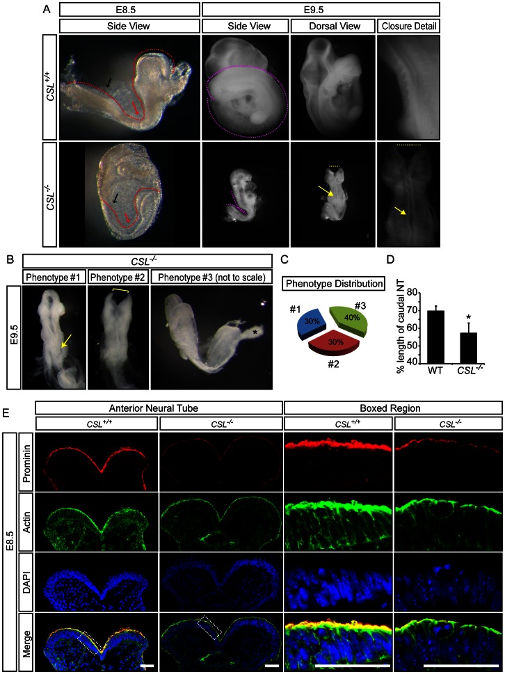
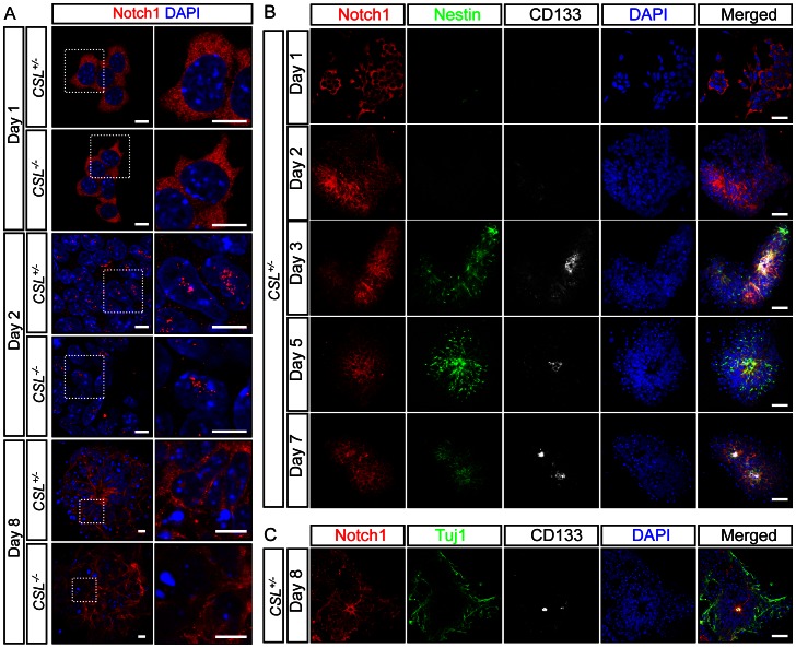
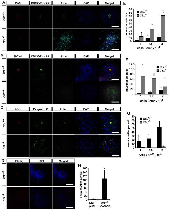
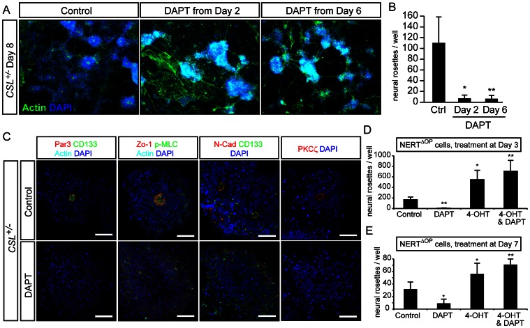
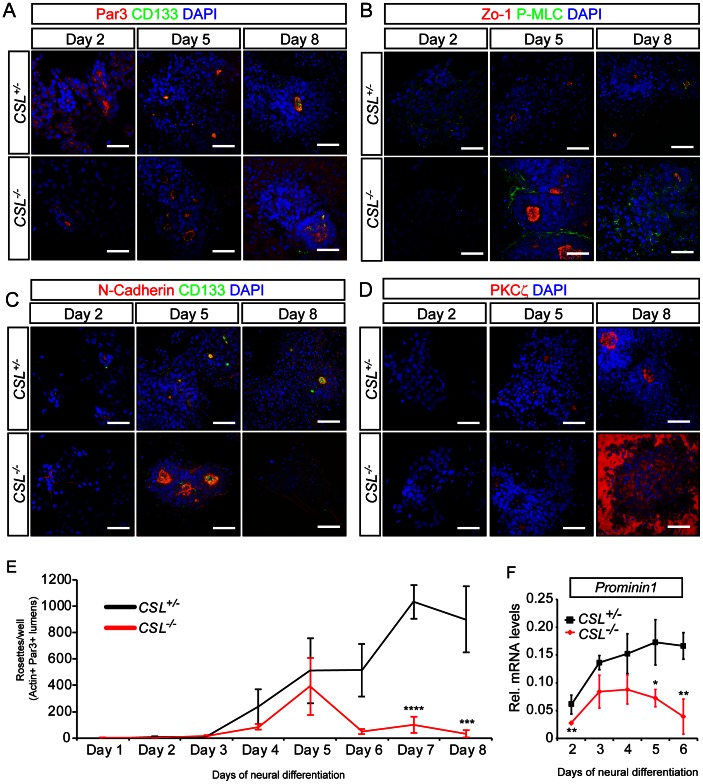
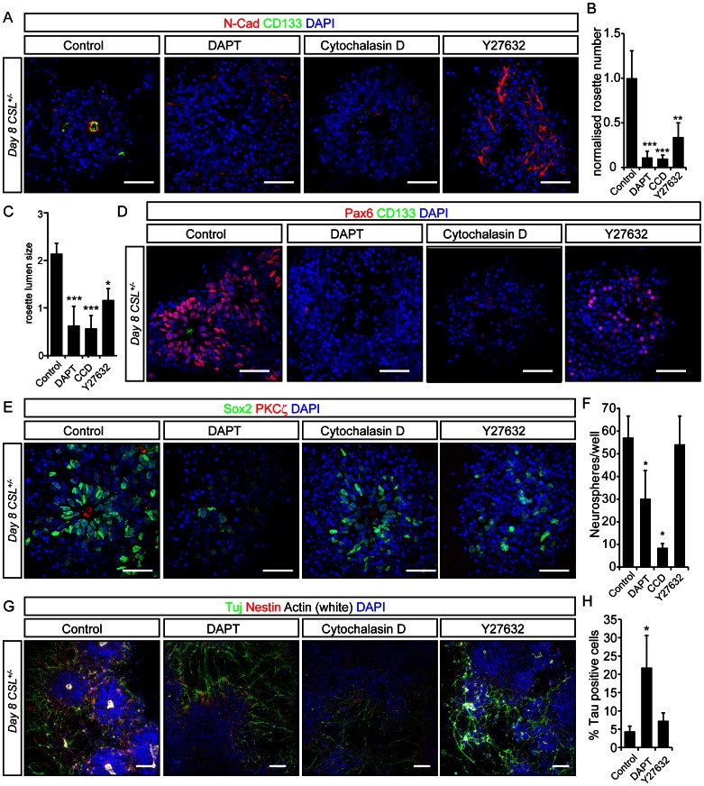
Formation of the metazoan body plan requires a complex interplay of morphological changes and patterning, and central to these processes is the establishment of apical/basal cell polarity. In the developing nervous system, apical/basal cell polarity is essential for neural tube closure and maintenance of the neural stem cell population. In this report we explore how a signaling pathway important for nervous system development, Notch signaling, impacts on apical/basal cell polarity in neural differentiation. CSL(-/-) mouse embryos, which are devoid of canonical Notch signaling, demonstrated a neural tube phenotype consistent with cell polarity and convergent extension defects, including deficiencies in the restricted expression of apical polarity markers in the neuroepithelium. CSL(-/-) mouse embryonic stem (ES) cells, cultured at low density, behaved as wild-type in the establishment of neural progenitors and apical specification, though progression through rosette formation, an in vitro correlate of neurulation, required CSL for correct maintenance of rosette structure and regulation of neuronal differentiation. Similarly, acute pharmacological inhibition of Notch signaling led to the breakdown of neural rosettes and accelerated neuronal differentiation. In addition to functional Notch signaling, rosette integrity was found to require actin polymerization and Rho kinase (ROCK) activity. Disruption of rosettes through inhibition of actin polymerization or ROCK activity, however, had no effect on neuronal differentiation, indicating that rosette maintenance is not a prerequisite for normal neuronal differentiation. In conclusion, our data indicate that Notch signaling plays a role not only in differentiation, but also in organization and maintenance of polarity during development of the early nervous system.


Conflict of interest statement

Competing Interests: The authors have declared that no competing interests exist.

Figures

Figure 1
Figure 1. Notch signalling is required for appropriate neurulation.
(A) At E8.5, CSL−/− embryos are somewhat developmentally delayed and display a shorter anterior-posterior axis (red dotted line) than WT littermates, which is particularly evident in the caudal region (black arrow). At E9.5, CSL−/− embryos are still smaller than wild-type littermates, and have usually failed to turn. CSL−/− embryos display neural tube defects such as an open anterior neural tube (yellow dotted lines), a kinked neural tube closure line in the thoracic levels (yellow arrow) and convergent extension defects with a shortened caudal anterior-posterior axis (pink dotted lines). (B, C) At E9.5, CSL−/− embryos are obtained at approximately Mendelian ratios (27.78%, 10 out of 36) and these manifest three different levels of penetrance of the phenotype, present in approximately equal frequencies (#1 = 30%, #2 = 30%, #3 = 40%). The mildest CSL−/− phenotype (#1) manifests as a slight developmental delay, disrupted caudal development and an undulating neural tube closure line (yellow arrow). Phenotype #2 includes an equivalent developmental delay as seen in Phenotype #1, but these embryos have an open neural tube in both the anterior (yellow bracket) and posterior regions. Phenotype #3 is grossly developmentally delayed, resembling approximately E8.5, and has not yet undergone neurulation. The images for wild-type, Phenotype #1 and #2 were taken at the same magnification, while Phenotype #3 was taken at a larger magnification as it was much smaller. (D) Convergent extension was quantified in E9 embryos by dividing the length of the caudal neural tube (from the caudal hindbrain to the tip of the tail), in which convergent extension is the most pronounced, by the length of the entire neural tube (from anterior forebrain to the tip of the tail). (WT N = 4, CSL−/− N = 3, p = 0.047). (E) Coronal sections of E8.5 anterior neural tube show decreased apical actin and CD133 staining in CSL−/− embryos. Scale bar is 50 μm. Please see Fig. S1 for additional markers at other stages.
Figure 2
Figure 2. Notch1 is expressed in ES cells undergoing neural differentiation.
(A) Notch1 is detected homogenously throughout the cytoplasm of ES cells undergoing neural differentiation at Day 1, using an antibody specific for the C-terminus of Notch1 (detecting cleaved and full-length Notch1). Notch1 translocates to the nucleus on Day 2 or 3 of neural differentiation, and can be found in two or three specific nuclear loci. Notch1 is detectable until Day 8. (B) During differentiation Notch1 expression is enriched in Nestin+ neural progenitor cells from Day 3, and is maintained until Day 8. (C) Notch1 expression does not coincide with Tuj+ neurons, shown here at Day 8. In (A) scale bar is 10 μm, in (B) and (C) scale bar is 40 μm. For separate channels please see Fig. S3.
Figure 3
Figure 3. Notch signalling is required for the presence of neural rosettes at Day 8 of differentiation.
(A) CSL+/− and CSL−/− ES cells were differentiated for 8 days under neural differentiation conditions. CSL−/− differentiations do not contain rosettes, as assessed by staining for rosette lumen-specific markers (A) Par3 (Pard3)/CD133 (Prominin)/Actin, (B) Zo-1/phosphorylated myosin light chain (P-MLC)/Actin, (C) N-Cadherin/CD133Actin, and (D) PKCξ stainings. (E–F) Seeding density (1, 1.5 and 2×10⁴ cells/cm2) contributes to the numbers of non-neural (E) and neuronal (F), colonies, per well, obtained from CSL−/− ES cells but no seeding density rescued rosette formation (G) after 8 days of neural differentiation. (H) The lack of rosettes in CSL−/− differentiations is CSL-specific, and not cell-line specific, as the rosette defect seen in CSL−/− ES cells can be rescued by stable re-introduction of CSL. Scale bars in all panels are 50 μm. For separate channels please see Fig. S4.
Figure 4
Figure 4. Notch is required for rosette maintenance.
(A, B) Day8 CSL+/− ES cell neural differentiations, treated with the γ-secretase inhibitor DAPT from Day 2 or Day 6, display a drastic reduction in rosette number, visualized with staining for actin and DAPI (A), quantified in (B). (C) Acute treatment of CSL+/− cells with DAPT for 16 hours between Day 7 and Day 8 leads to a break-down of existing rosettes, as assessed by Par3 (Pard3)/CD133 (Prominin)/Actin, Zo-1/phosphorylated myosin light chain (P-MLC)/Actin, N-Cadherin (N-Cad)/CD133/Actin, and PKCξ stainings. Remnants of rosettes, cells organized in rings, can be found instead. (D,E) Another ES cell line (NERTΔOP), with tamoxifen-inducible Notch1 signalling, confirms that repression of Notch signalling with DAPT reduces rosette numbers, and also reveals that activation of Notch signalling in the presence or absence of DAPT up-regulates the number of rosettes, whether Notch activity is induced on day 3 (D) or day 7 (E). Images in (A) were acquired on a fluorescence microscope at 10× magnification. Scale bars in (C) are 50 μm. Bar graphs depict means from three experiments performed in triplicate, error bars indicate standard deviation: ***significant difference at p<0.001; **significant difference at p<0.01; *significant difference at p<0.05. For separate channels please see Fig. S6.
Figure 5
Figure 5. Notch signalling is not required for the acquisition of polarity and initial development of rosettes during neural differentiation.
Immunohistochemistry for (A) Par3/CD133, (B) Zo-1/phosphorylated myosin light chain (PMLC), (C) N-cadherin/CD133 and (D) PKCζ reveal that up until day 5, lumen/polarity stains are similar in CSL+/− and CSL−/− differentiations, and that CSL−/− differentiations display lumens at around day 5, similar to CSL+/− differentiations. The development of these lumens is quantified in (E), showing that indeed development is similar up to day 5 (N = 3). (F) CD133 qPCR follows a similar pattern over 6 days of differentiation, with an initially normal induction and subsequent loss of CD133 mRNA. Error bars show standard deviation. ***significant difference at p<0.001; **significant difference at p<0.01; *significant difference at p<0.05. All scale bars are 50 μm.
Figure 6
Figure 6. Loss of Notch signaling accelerates neuronal differentiation.
Time-course analysis of Pax6 (A) and Sox1 (B) mRNA in CSL+/− and CSL−/− ES cell neural differentiations reveal normal neural induction but a rapid depletion of neuroepithelial progenitors in CSL−/− differentiations. Immunohistochemistry for Pax6, Tuj1 (C) and Sox2 (D) confirm that these markers are induced lost beginning at day 5 in CSL−/− differentiations. Note that oval structures with neural stem cells surrounding a central lumen (yellow arrows) can be found in both CSL+/− and CSL−/− cultures at day 5. (E,F) A larger number of neurospheres are derived from CSL+/− cultures after 7 days of monolayer differentiation than from CSL−/− differentiations, quantified in (F). In addition, CSL−/− spheres display an aberrant appearance with cells bulging out (boxed region in E). At day 8 (G) the neural stem cell marker Sox3 and the radial glia marker and Notch target gene BLBP are absent in CSL−/− differentiations. BLBP mRNA (H) is not detected in CSL−/− cultures over 6 days of differentiation. Finally, CSL−/− differentiations (I) lose Nestin expression and differentiate into Tuj1+ neurons more rapidly than CSL+/− differentiations, this is statistically significant (J) from day 3 of differentiation, as seen in this quantification of the number of colonies in CSL+/− and CSL−/− differentiations containing TuJ1 positive cells at day 2 and day 3. Scalebar in (C) (confocal, 5 μm section, 20×) and (D) (confocal, 2 μm section, 40×) is 100 μm, scalebar in (G) (confocal, 2 μm section, 20×) is 50 μm. Panels in (I) were taken on a fluorescence microscope at 10×. Graphs depict means from three experiments performed in triplicate, error bars indicate standard deviation: ***significant difference at p<0.001; **significant difference at p<0.01; *significant difference at p<0.05.
Figure 7
Figure 7. Differentiation of neural rosettes into neurons is not dependent on appropriate polarity cues in vitro.
(A) Immunohistochemistry for N-Cadherin and CD133, of CSL+/− rosettes treated with the γ-secretase inhibitor DAPT, Cytochalasin D or Y27632 overnight between day 7 and day 8 depletes rosettes, leaving rings of cells lacking apical markers. Loss of rosette structures 15 hours after initiation of treatment is quantified in (B). Measure of lumen size 12 hours after initiation of treatment demonstrates a reduction in lumen size, quantified in (C). Neural stem cell markers Pax6 (D) and Sox2 (E) are dramatically reduced by DAPT treatment, but are less severely affected by Cytochalasin D or Y27632. (F) Neurospheres derived from CSL+/− cultures treated overnight between day 7 and 8 reveal that DAPT treatment and Cytochalasin D compromises neural stem cell potential, while Y27632 has no effect on stem cell potential. (G) Immunohistochemistry for Tuj1, Nestin and phalloidin staining for actin shows that DAPT redistributes actin and induces neuronal differentiation, while Y27632 and Cytochalasin D redistribute actin staining but have no discernible effect on TuJ1+ neurons. (H) The effect of loss of polarity on neuronal differentiation was quantified using a Tau-GFP ES cell line using FACS. Only DAPT induced an increase in the number of neurons 48 hours after initiation of inhibitor treatment. Please see Fig. S15 for gating. Bar graphs depict means from three experiments performed in triplicate, error bars indicate standard deviation: ***significant difference at p<0.001; **significant difference at p<0.01; *significant difference at p<0.05.

References

    1. Copp AJ, Greene ND, Murdoch JN (2003) The genetic basis of mammalian neurulation. Nat Rev Genet 4: 784–793. - PubMed
    1. Mitchell KJ (2011) The genetics of neurodevelopmental disease. Curr Opin Neurobiol 21: 197–203. - PubMed
    1. Doudney K, Stanier P (2005) Epithelial cell polarity genes are required for neural tube closure. Am J Med Genet C Semin Med Genet 135C: 42–47. - PubMed
    1. Desmond ME, Schoenwolf GC (1986) Evaluation of the roles of intrinsic and extrinsic factors in occlusion of the spinal neurocoel during rapid brain enlargement in the chick embryo. J Embryol Exp Morphol 97: 25–46. - PubMed
    1. Ybot-Gonzalez P, Savery D, Gerrelli D, Signore M, Mitchell CE, et al. (2007) Convergent extension, planar-cell-polarity signalling and initiation of mouse neural tube closure. Development 134: 789–799. - PMC - PubMed

Publication types

MeSH terms

LinkOut - more resources