

Mice lacking pten in osteoblasts have improved intramembranous and late endochondral fracture healing
- PMID: 23675511
- PMCID: PMC3652860
- DOI: 10.1371/journal.pone.0063857
Mice lacking pten in osteoblasts have improved intramembranous and late endochondral fracture healing
Abstract
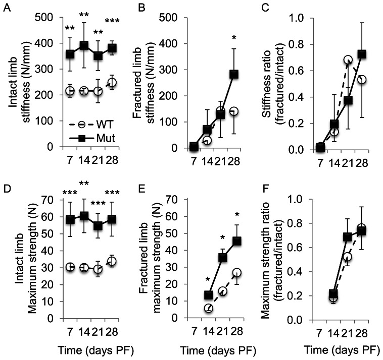
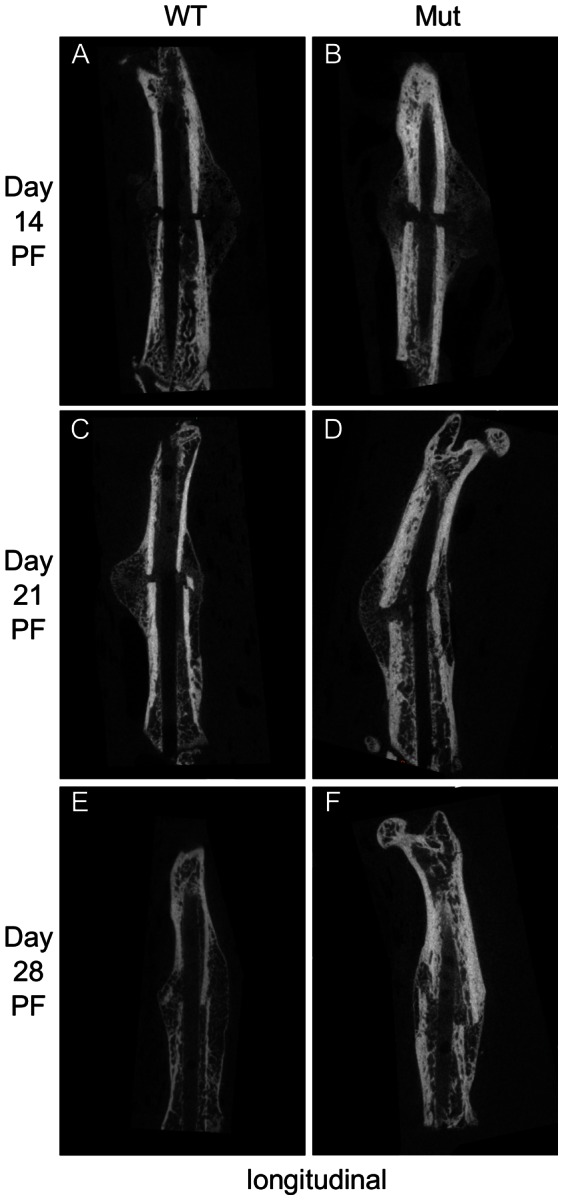
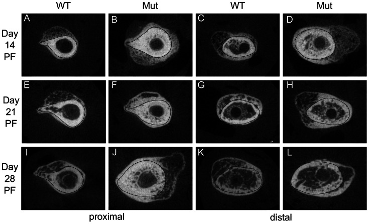
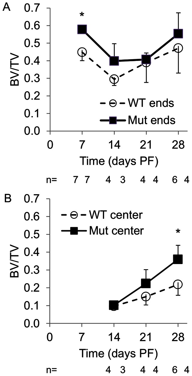
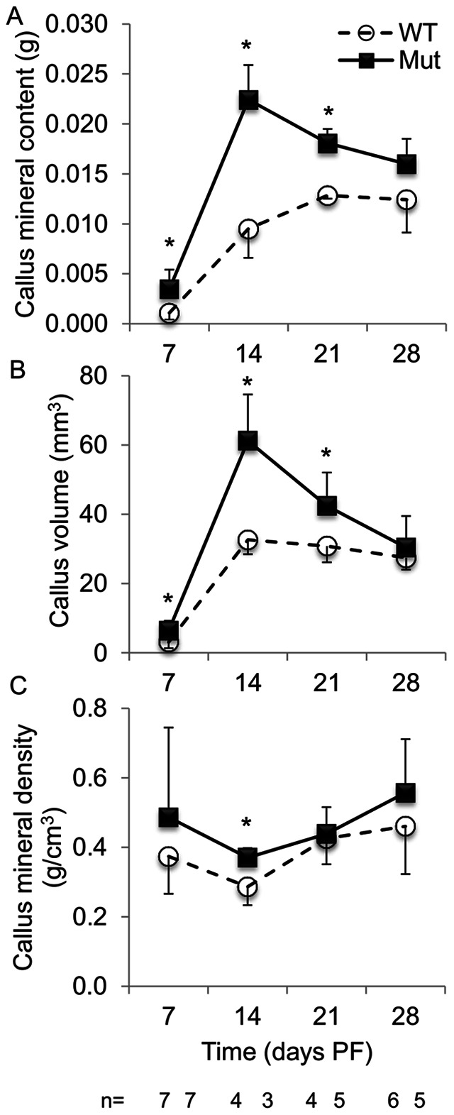
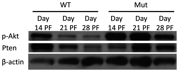
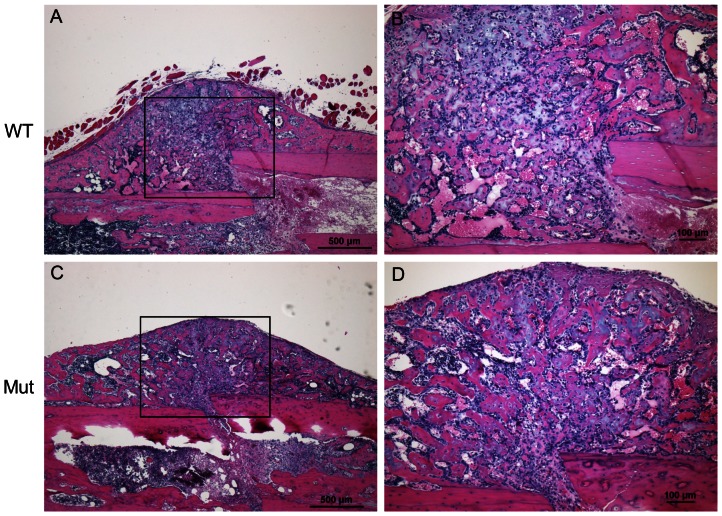
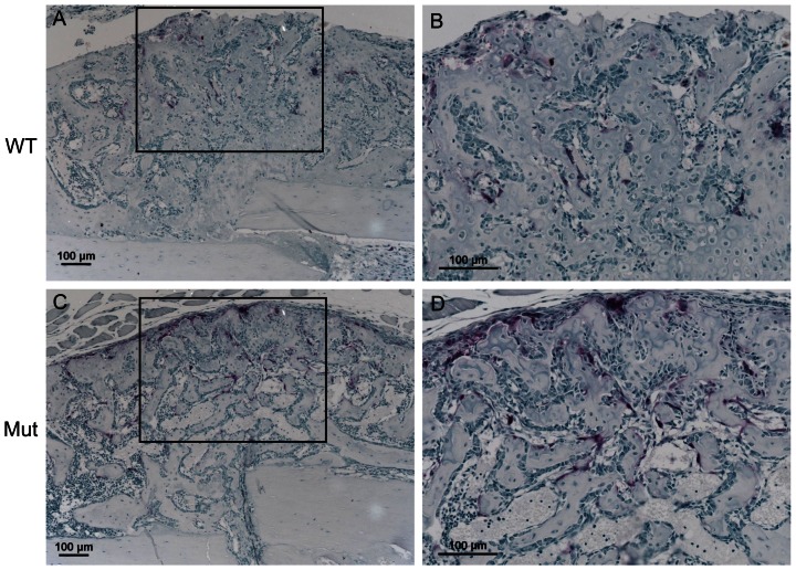
The failure of an osseous fracture to heal (development of a non-union) is a common and debilitating clinical problem. Mice lacking the tumor suppressor Pten in osteoblasts have dramatic and progressive increases in bone volume and density throughout life. Since fracture healing is a recapitulation of bone development, we investigated the process of fracture healing in mice lacking Pten in osteoblasts (Ocn-cre(tg/+;)Pten(flox/flox) ). Mid-diaphyseal femoral fractures induced in wild-type and Ocn-cre(tg/+;)Pten(flox/flox) mice were studied via micro-computed tomography (µCT) scans, biomechanical testing, histological and histomorphometric analysis, and protein expression analysis. Ocn-cre(tg/+;)Pten(flox/flox) mice had significantly stiffer and stronger intact bones relative to controls in all cohorts. They also had significantly stiffer healing bones at day 28 post-fracture (PF) and significantly stronger healing bones at days 14, 21, and 28 PF. At day 7 PF, the proximal and distal ends of the Pten mutant calluses were more ossified. By day 28 PF, Pten mutants had larger and more mineralized calluses. Pten mutants had improved intramembranous bone formation during healing originating from the periosteum. They also had improved endochondral bone formation later in the healing process, after mature osteoblasts are present in the callus. Our results indicate that the inhibition of Pten can improve fracture healing and that the local or short-term use of commercially available Pten-inhibiting agents may have clinical application for enhancing fracture healing.
Conflict of interest statement
Figures
References
-
- Phillips AM (2005) Overview of the fracture healing cascade. Injury 36 Suppl 3S5–7. - PubMed
-
- Ferguson CM, Miclau T, Hu D, Alpern E, Helms JA (1998) Common molecular pathways in skeletal morphogenesis and repair. Ann N Y Acad Sci 857: 33–42. - PubMed
-
- Einhorn TA (1998) The cell and molecular biology of fracture healing. Clin Orthop Relat Res: S7–21. - PubMed
-
- Gerstenfeld LC, Cullinane DM, Barnes GL, Graves DT, Einhorn TA (2003) Fracture healing as a post-natal developmental process: molecular, spatial, and temporal aspects of its regulation. J Cell Biochem 88: 873–884. - PubMed
-
- Parker MJ, Raghavan R, Gurusamy K (2007) Incidence of fracture-healing complications after femoral neck fractures. Clin Orthop Relat Res 458: 175–179. - PubMed
Publication types
MeSH terms
Substances
Grants and funding
LinkOut - more resources
Full Text Sources
Other Literature Sources
Molecular Biology Databases
Research Materials
Miscellaneous
