

Generation and characterization of an Nxf7 knockout mouse to study NXF5 deficiency in a patient with intellectual disability
- PMID: 23675524
- PMCID: PMC3652825
- DOI: 10.1371/journal.pone.0064144
Generation and characterization of an Nxf7 knockout mouse to study NXF5 deficiency in a patient with intellectual disability
Abstract
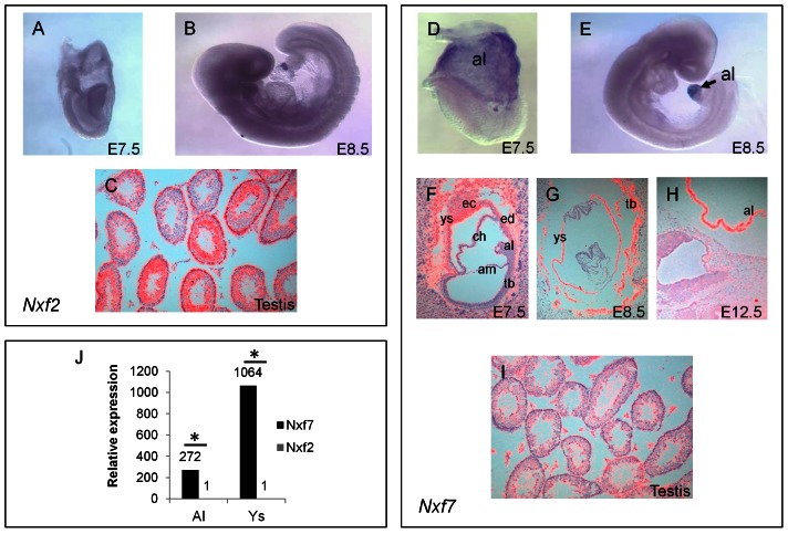
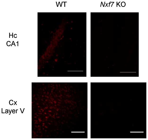
Members of the Nuclear eXport Factor (NXF) family are involved in the export of mRNA from the nucleus to the cytoplasm, or hypothesized to play a role in transport of cytoplasmic mRNA. We previously reported on the loss of NXF5 in a male patient with a syndromic form of intellectual disability. To study the functional role of NXF5 we identified the mouse counterpart. Based on synteny, mouse Nxf2 is the ortholog of human NXF5. However, we provide several lines of evidence that mouse Nxf7 is the actual functional equivalent of NXF5. Both Nxf7 and NXF5 are predominantly expressed in the brain, show cytoplasmic localization, and present as granules in neuronal dendrites suggesting a role in cytoplasmic mRNA metabolism in neurons. Nxf7 was primarily detected in the pyramidal cells of the hippocampus and in layer V of the cortex. Similar to human NXF2, mouse Nxf2 is highly expressed in testis and shows a nuclear localization. Interestingly, these findings point to a different evolutionary path for both NXF genes in human and mouse. We thus generated and validated Nxf7 knockout mice, which were fertile and did not present any gross anatomical or morphological abnormalities. Expression profiling in the hippocampus and the cortex did not reveal significant changes between wild-type and Nxf7 knockout mice. However, impaired spatial memory was observed in these KO mice when evaluated in the Morris water maze test. In conclusion, our findings provide strong evidence that mouse Nxf7 is the functional counterpart of human NXF5, which might play a critical role in mRNA metabolism in the brain.
Conflict of interest statement
Figures


Similar articles
-
Nxf7 deficiency impairs social exploration and spatio-cognitive abilities as well as hippocampal synaptic plasticity in mice.Front Behav Neurosci. 2015 Jul 10;9:179. doi: 10.3389/fnbeh.2015.00179. eCollection 2015. Front Behav Neurosci. 2015. PMID: 26217206 Free PMC article.
-
NXF5, a novel member of the nuclear RNA export factor family, is lost in a male patient with a syndromic form of mental retardation.Curr Biol. 2001 Sep 18;11(18):1381-91. doi: 10.1016/s0960-9822(01)00419-5. Curr Biol. 2001. PMID: 11566096
-
Molecular cloning and functional characterization of mouse Nxf family gene products.Genomics. 2005 May;85(5):641-53. doi: 10.1016/j.ygeno.2005.01.003. Genomics. 2005. PMID: 15820316
-
NXF2 is involved in cytoplasmic mRNA dynamics through interactions with motor proteins.Nucleic Acids Res. 2007;35(8):2513-21. doi: 10.1093/nar/gkm125. Epub 2007 Apr 1. Nucleic Acids Res. 2007. PMID: 17403691 Free PMC article.
-
RNA-binding proteins of the NXF (nuclear export factor) family and their connection with the cytoskeleton.Cytoskeleton (Hoboken). 2017 Apr;74(4):161-169. doi: 10.1002/cm.21362. Epub 2017 Apr 1. Cytoskeleton (Hoboken). 2017. PMID: 28296067 Review.
Cited by
-
A Drosophila model system to assess the function of human monogenic podocyte mutations that cause nephrotic syndrome.Hum Mol Genet. 2017 Feb 15;26(4):768-780. doi: 10.1093/hmg/ddw428. Hum Mol Genet. 2017. PMID: 28164240 Free PMC article.
-
Nxf7 deficiency impairs social exploration and spatio-cognitive abilities as well as hippocampal synaptic plasticity in mice.Front Behav Neurosci. 2015 Jul 10;9:179. doi: 10.3389/fnbeh.2015.00179. eCollection 2015. Front Behav Neurosci. 2015. PMID: 26217206 Free PMC article.
-
A Patient with a Small Deletion Affecting Only Exon 1-Intron 1 of the NXF5 Gene: Potential Evidence Supporting Its Role in Neurodevelopmental Disorders.Genes (Basel). 2025 May 13;16(5):571. doi: 10.3390/genes16050571. Genes (Basel). 2025. PMID: 40428393 Free PMC article.
-
Respiratory failure, cleft palate and epilepsy in the mouse model of human Xq22.1 deletion syndrome.Hum Mol Genet. 2014 Jul 15;23(14):3823-9. doi: 10.1093/hmg/ddu095. Epub 2014 Feb 25. Hum Mol Genet. 2014. PMID: 24569167 Free PMC article.
-
Sending the message: specialized RNA export mechanisms in trypanosomes.Trends Parasitol. 2022 Oct;38(10):854-867. doi: 10.1016/j.pt.2022.07.008. Epub 2022 Aug 24. Trends Parasitol. 2022. PMID: 36028415 Free PMC article. Review.
References
-
- Jun L, Frints SGM, Duhamel H, Herold A, Abad-Rodrigues J, et al. (2001) NXF5, a novel member of the nuclear RNA export factor family, is lost in a male patient with a syndromic form of mental retardation. Curr Biol 11: 1381–1391. - PubMed
-
- Yang J, Bogerd HP, Wang PJ, Page DC, Cullen BR (2001) Two closely related human nuclear export factors utilize entirely distinct export pathways. Mol Cell 8: 397–406. - PubMed
Publication types
MeSH terms
Substances
LinkOut - more resources
Full Text Sources
Other Literature Sources
Molecular Biology Databases
Research Materials
