

Genomic approach to identify factors that drive the formation of three-dimensional structures by EA.hy926 endothelial cells
- PMID: 23675535
- PMCID: PMC3651237
- DOI: 10.1371/journal.pone.0064402
Genomic approach to identify factors that drive the formation of three-dimensional structures by EA.hy926 endothelial cells
Abstract
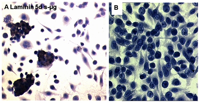
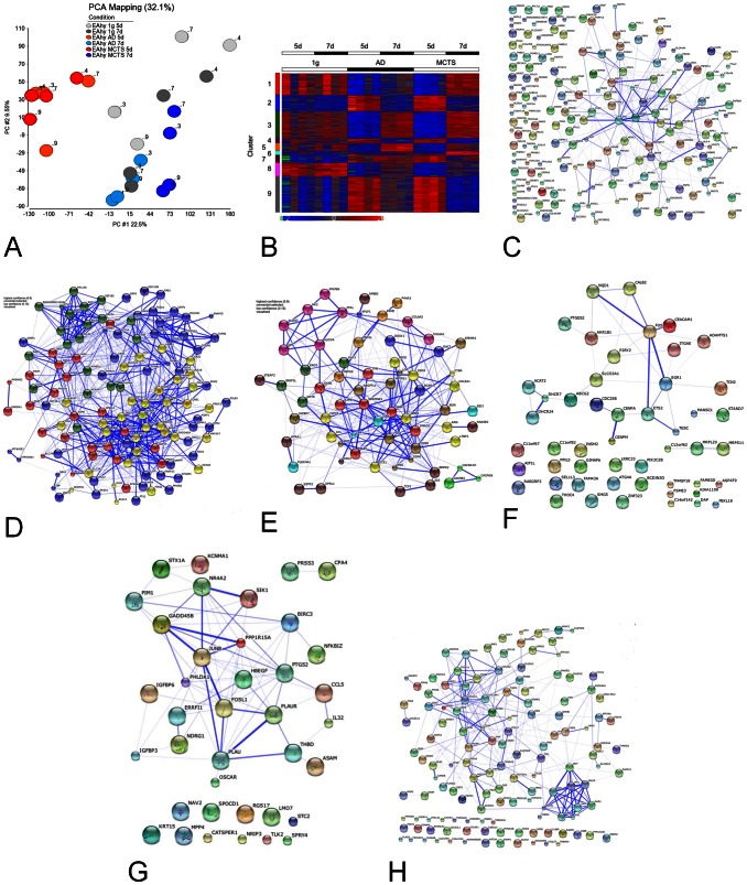
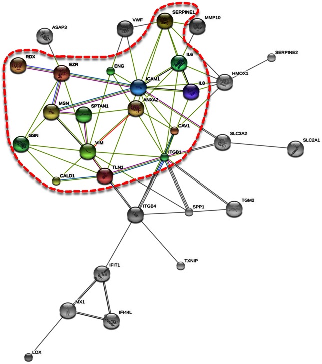
Understanding the mechanisms responsible for tube formation by endothelial cells (ECs) is of major interest and importance in medicine and tissue engineering. Endothelial cells of the human cell line EA.hy926 behave ambivalently when cultured on a random positioning machine (RPM) simulating microgravity. Some cells form tube-like three-dimensional (3D) aggregates, while other cells (AD) continue to grow adherently. Between the fifth and seventh day of culturing, the two types of cell growth achieve the greatest balance. We harvested ECs that grew either adherently or as 3D aggregates separately after five and seven days of incubation on the RPM, and applied gene array analysis and PCR techniques to investigate their gene expression profiles in comparison to ECs growing adherently under normal static 1 g laboratory conditions for equal periods of time. Using gene arrays, 1,625 differentially expressed genes were identified. A strong overrepresentation of transient expression differences was found in the five-day, RPM-treated samples, where the number of genes being differentially expressed in comparison to 1 g cells was highest as well as the degree of alteration regarding distinct genes. We found 27 genes whose levels of expression were changed at least 4-fold in RPM-treated cells as compared to 1 g controls. These genes code for signal transduction and angiogenic factors, cell adhesion, membrane transport proteins or enzymes involved in serine biosynthesis. Fifteen of them, with IL8 (interleukin 8) and VWF (von Willebrand factor) the most prominently affected, showed linkages to genes of another 20 proteins that are important in cell structure maintenance and angiogenesis and extended their network of interaction. Thus, the study reveals numerous genes, which mutually influence each other during initiation of 3D growth of endothelial cells.
Conflict of interest statement
Figures
Similar articles
-
Proteomic differences between microvascular endothelial cells and the EA.hy926 cell line forming three-dimensional structures.Proteomics. 2014 Mar;14(6):689-98. doi: 10.1002/pmic.201300453. Epub 2014 Feb 20. Proteomics. 2014. PMID: 24376074
-
Key Proteins Involved in Spheroid Formation and Angiogenesis in Endothelial Cells After Long-Term Exposure to Simulated Microgravity.Cell Physiol Biochem. 2018;45(2):429-445. doi: 10.1159/000486920. Epub 2018 Jan 24. Cell Physiol Biochem. 2018. PMID: 29402845
-
Growth of Endothelial Cells in Space and in Simulated Microgravity - a Comparison on the Secretory Level.Cell Physiol Biochem. 2019;52(5):1039-1060. doi: 10.33594/000000071. Cell Physiol Biochem. 2019. PMID: 30977987
-
Scaffold-free Tissue Formation Under Real and Simulated Microgravity Conditions.Basic Clin Pharmacol Toxicol. 2016 Oct;119 Suppl 3:26-33. doi: 10.1111/bcpt.12561. Epub 2016 Feb 29. Basic Clin Pharmacol Toxicol. 2016. PMID: 26826674 Review.
-
Branching out: a molecular fingerprint of endothelial differentiation into tube-like structures generated by Affymetrix oligonucleotide arrays.Microcirculation. 2003 Jan;10(1):63-81. doi: 10.1038/sj.mn.7800170. Microcirculation. 2003. PMID: 12610664 Review.
Cited by
-
The impact of microgravity and hypergravity on endothelial cells.Biomed Res Int. 2015;2015:434803. doi: 10.1155/2015/434803. Epub 2015 Jan 13. Biomed Res Int. 2015. PMID: 25654101 Free PMC article. Review.
-
Microgravity and Space Medicine 2.0.Int J Mol Sci. 2022 Apr 18;23(8):4456. doi: 10.3390/ijms23084456. Int J Mol Sci. 2022. PMID: 35457274 Free PMC article.
-
Structural and Molecular Changes of Human Chondrocytes Exposed to the Rotating Wall Vessel Bioreactor.Biomolecules. 2023 Dec 24;14(1):25. doi: 10.3390/biom14010025. Biomolecules. 2023. PMID: 38254625 Free PMC article.
-
Thyroid cancer cells in space during the TEXUS-53 sounding rocket mission - The THYROID Project.Sci Rep. 2018 Jul 9;8(1):10355. doi: 10.1038/s41598-018-28695-1. Sci Rep. 2018. PMID: 29985426 Free PMC article.
-
Recent Advances in Breast Cancer Research.Int J Mol Sci. 2023 Jul 26;24(15):11990. doi: 10.3390/ijms241511990. Int J Mol Sci. 2023. PMID: 37569366 Free PMC article.
References
-
- Cines DB, Pollak ES, Buck CA, Loscalzo J, Zimmerman GA, et al. (1998) Endothelial cells in physiology and in the pathophysiology of vascular disorders. Blood 91: 3527–3561. - PubMed
-
- Arnold F, West DC (1991) Angiogenesis in wound healing. Pharmacol Ther 52: 407–422. - PubMed
-
- Reynolds LP, Redmer DA (2001) Angiogenesis in the placenta. Biol Reprod 64: 1033–1040. - PubMed
-
- Folkman J (1995) Angiogenesis in cancer, vascular, rheumatoid and other disease. Nature Med 1: 27–31. - PubMed
Publication types
MeSH terms
LinkOut - more resources
Full Text Sources
Other Literature Sources
Miscellaneous
