. 2013 May 15:11:43.
doi: 10.1186/1477-7827-11-43.

Intraovarian regulation of gonadotropin-dependent folliculogenesis depends on notch receptor signaling pathways not involving Delta-like ligand 4 (Dll4)

Affiliations

Intraovarian regulation of gonadotropin-dependent folliculogenesis depends on notch receptor signaling pathways not involving Delta-like ligand 4 (Dll4)

Vuk P Jovanovic et al. Reprod Biol Endocrinol. .

Abstract

Background: In-situ hybridisation studies demonstrate that Notch receptors and ligands are expressed in granulosa cells (GCs) and in the theca layer vasculature of growing follicles. Notch signaling involves cell-to-cell interaction mediated by transmembrane receptors and ligands. This signaling pathway may represent a novel intraovarian regulator of gonadotropin-dependent follicular development to the preovulatory stage. We hypothesized that blocking Notch pathways would disrupt follicular maturation in the mouse ovary.

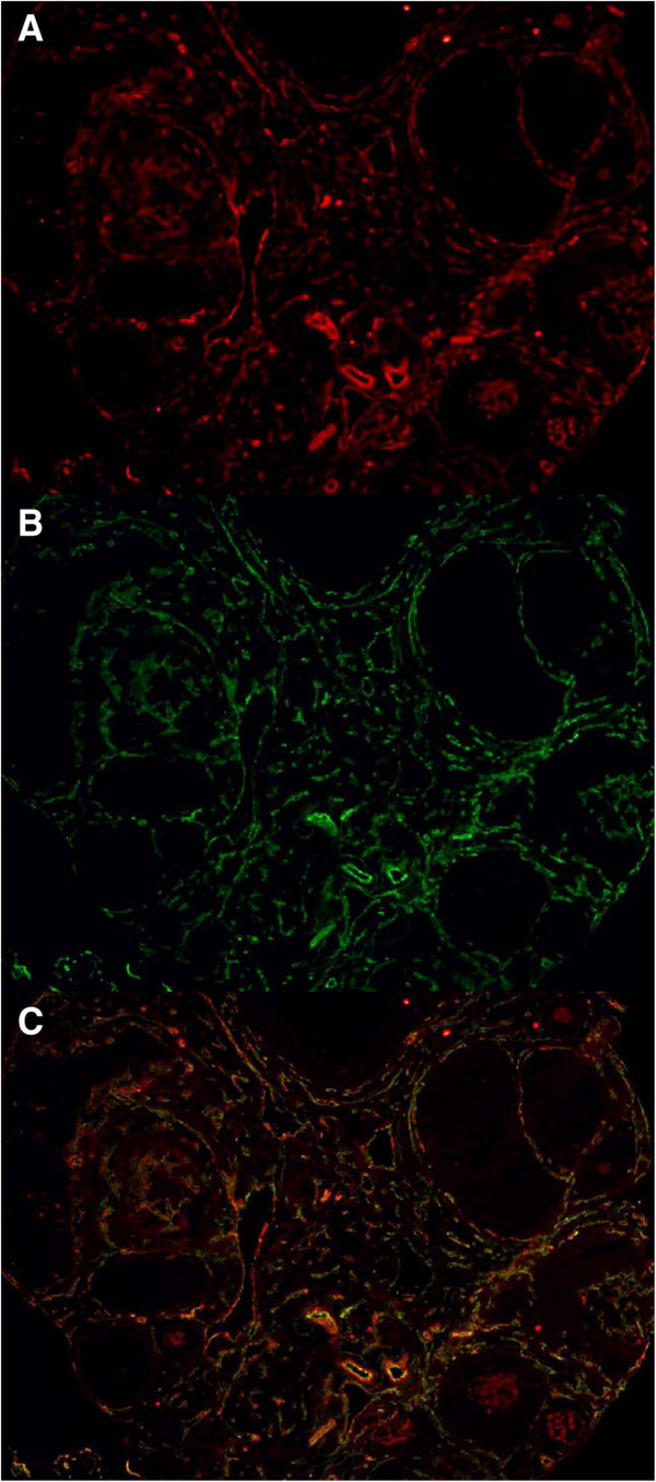
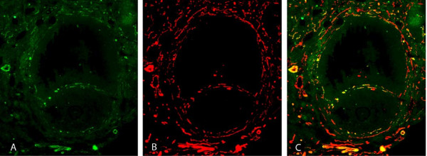
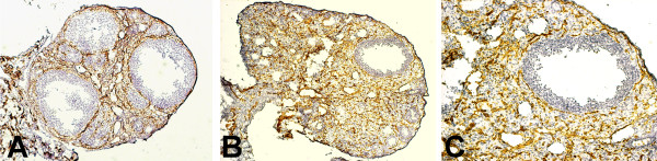
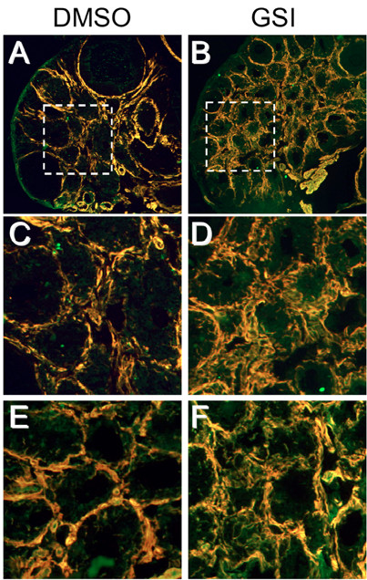
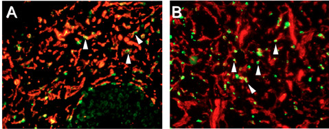
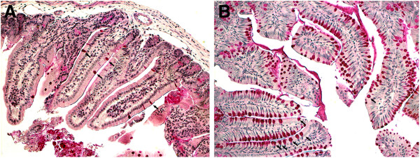
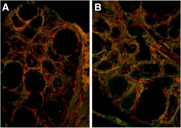
Methods: Hypophysectomized CD21 female mice were administered pregnant mare serum gonadotropin (PMSG) for 3 days to stimulate follicular development. In one experiment, a pan-notch inhibitor, compound E, was initiated 2 days prior to and throughout stimulation (n = 10), while in a second experiment, a humanized phage Dll4 blocking antibody, YW152F, was used (n = 5). After sacrifice, ovarian histology, serum estradiol levels and uterine weights were compared to controls. The ovarian morphology was evaluated with hematoxylin/eosin staining and immunohistochemistry was performed for Notch1, Notch2, Notch3, Notch4, Jagged1, Dll4, platelet endothelial cell adhesion molecule (PECAM) and alpha-smooth muscle actin (α-SMA) detection.

Results: We localized specific Notch ligands and receptors in the following structures: Dll4 is specific to theca layer endothelial cells (ECs); Notch1/Notch4 and Jagged1 are expressed in theca layer ECs and vascular smooth muscle cells (VSMCs), whereas Notch3 is restricted to VSMCs; Notch2 is expressed mostly on GCs of small follicles. Administration of a pan-Notch inhibitor, compound E, inhibits follicular development to the preovulatory stage (8.5 preovulatory follicles in treatment vs. 3.4 preovulatory follicles in control, p < 0.01; average number per ovary) with significant secondary effects on ovarian and uterine weight and estradiol secretion in a setting of uninhibited vascular proliferation, but disorganized appearance of ECs and VSMCs. Inhibition of endothelial Notch1 function through the inactivation of its ligand Dll4 with the blocking antibody YW152F induces mild disorganisation of follicular vasculature, but has no significant effect on gonadotropin-dependent folliculogenesis.

Conclusions: Our experiments suggest that the complete blockage of the Notch signaling pathway with compound E impairs folliculogenesis and induces disruption of gonadotropin stimulated angiogenesis. It seems the mechanism involves Notch1 and Notch3, specifically, causing the improper assembly of ECs and VSMCs in the theca layer, although the potential role of non-angiogenic Notch signaling, such as Jagged2 to Notch2 in GCs, remains to be elucidated.


Figures

Figure 1
Figure 1
Notch2 (red) in developing follicles. Intense staining in secondary follicles and sporadic red staining in tertiary follicles with complete absence of staining in the peripheral theca layer of the depicted 2 large follicles (10× magnification, DAPI counterstaining in blue).
Figure 2
Figure 2
PMSG stimulated untreated ovary with tertiary follicles: A) Notch3 in red B) PECAM in green C) Composite image with overlapping areas in yellow. Please note presence of Notch3 staining in the peripheral theca layer and absence of Notch3 staining on granulosa cells. (10× magnification).
Figure 3
Figure 3
Doublestaining of a developing tertiary follicle: A) Dll4 shown in green B) PECAM in red C) composite overlapping images showing presence of Dll4 in endothelial cells of the theca vasculature layer.
Figure 4
Figure 4
Inhibition of Notch signaling with a γ-secretase inhibitor (Compound E) blocked follicle development to the preovulatory stage. Immunohistochemistry for PECAM (brown) counterstained with hematoxylin. Please note disorganized appearance of PECAM staining in treatment group. A) three control preovulatory follicles (10× magnification) B) γ-secretase inhibitor treated follicles (10× magnification) C) Enlargement of treatment follicle in B (20 × magnification).
Figure 5
Figure 5
Compound E inhibition of Notch signaling increased vascular smooth muscle cells (VSMC) density and altered VSMC organization in the ovarian stroma. A-D) Immunofluorescence staining (10× magnified) for Notch3 (green) and α-SMA (red). C, D) enlargement of boxes areas in upper panels (20× magnified). Please note increased thickness and disorganized appearance of VSMCs in B and D. E, F) PECAM (green) and α-SMA (red). Please note disorganisation and failure to overlap in F (20× magnified).
Figure 6
Figure 6
Cell proliferation. PECAM (red) and BrdU (green). A) DMSO control treated B) Compound E, (20 × magnification). Please note that proliferation was not blocked by compound E (B) and even might be increased over control.
Figure 7
Figure 7
Intestinal section: A) control (DMSO), B) treatment (compound E). Goblet cells in red. Please note the increase of intestinal goblet cells in B over A (20× magnification).
Figure 8
Figure 8
Control and Dll4 blocking antibody YW152F treated ovaries stimulated with PMSG (10× magnification). Doublestaining with PECAM (red) and a-SMA (green). A) Control ovary. B) YW152F treated ovary. Please note that vascular appearance is slightly more disorganized in B.

Similar articles

Cited by

References

    1. Palermo R. Differential actions of FSH and LH during folliculogenesis. Reprod Biomed Online. 2007;15:326–337. doi: 10.1016/S1472-6483(10)60347-1. - DOI - PubMed
    1. Johnson J, Espinoza T, McGaughey RW, Rawls A, Wilson-Rawls J. Notch pathway genes are expressed in mammalian ovarian follicles. Mech Dev. 2001;109:355–361. doi: 10.1016/S0925-4773(01)00523-8. - DOI - PubMed
    1. Zhang CP, Yang JL, Zhang J, Li L, Huang L, Ji SY, Hu ZY, Gao F, Liu YX. Notch signaling is involved in ovarian follicle development by regulating granulosa cell proliferation. Endocrinology. 2011;152:2437–2447. doi: 10.1210/en.2010-1182. - DOI - PubMed
    1. Vorontchikhina MA, Zimmermann RC, Shawber CJ, Tang H, Kitajewski J. Unique patterns of Notch1, Notch4 and Jagged1 expression in ovarian vessels during folliculogenesis and corpus luteum formation. Gene Expr Patterns. 2005;5:701–709. doi: 10.1016/j.modgep.2005.02.001. - DOI - PubMed
    1. Mailhos C, Modlich U, Lewis J, Harris A, Bicknell R, Ish-Horowicz D. Delta4, an endothelial specific notch ligand expressed at sites of physiological and tumor angiogenesis. Differentiation. 2001;69:135–144. doi: 10.1046/j.1432-0436.2001.690207.x. - DOI - PubMed

MeSH terms

LinkOut - more resources