. 2013 Jun;123(6):2604-15.
doi: 10.1172/JCI67008. Epub 2013 May 15.

PD-L1 blockade synergizes with IL-2 therapy in reinvigorating exhausted T cells

Affiliations

PD-L1 blockade synergizes with IL-2 therapy in reinvigorating exhausted T cells

Erin E West et al. J Clin Invest. 2013 Jun.

Abstract

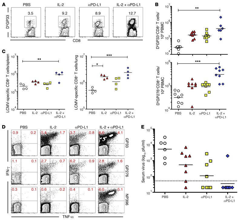
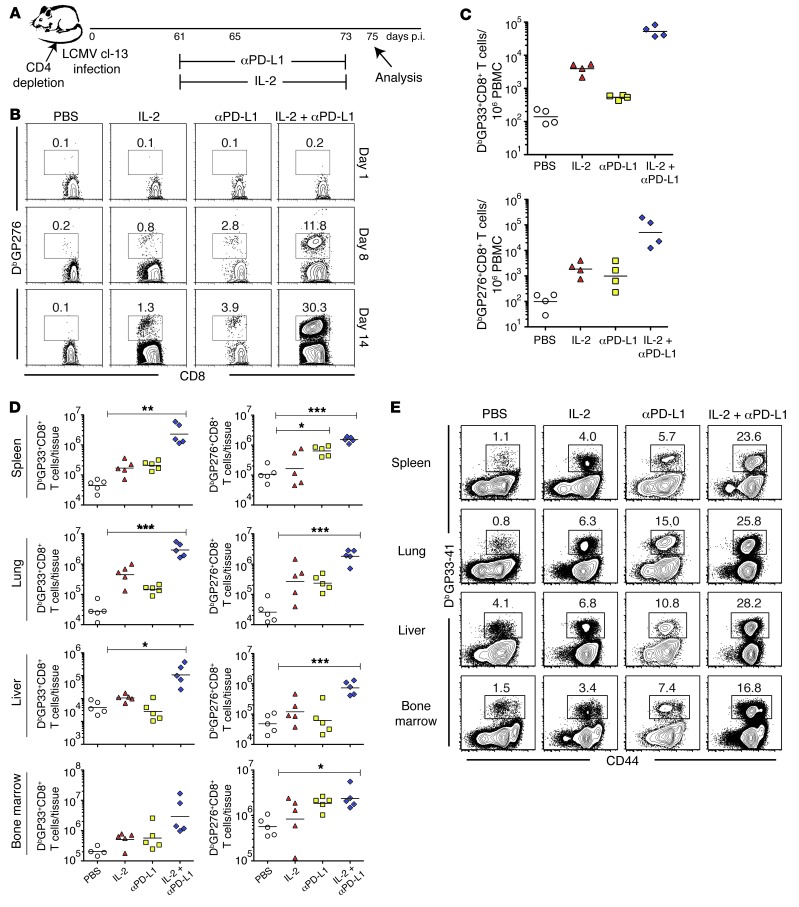
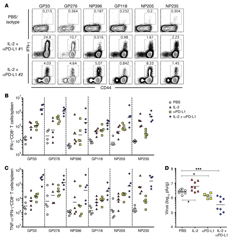
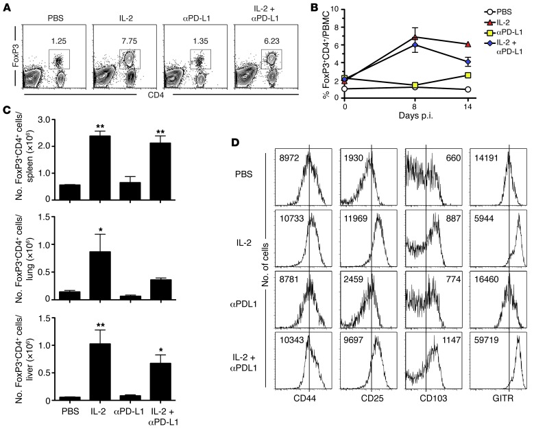
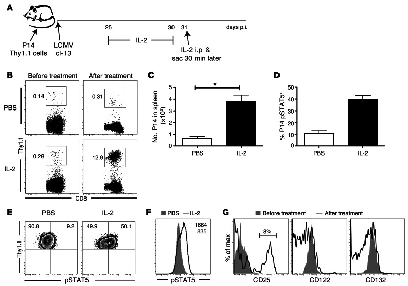
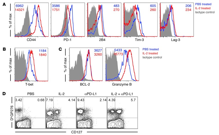
The inhibitory receptor programmed cell death 1 (PD-1) plays a major role in functional exhaustion of T cells during chronic infections and cancer, and recent clinical data suggest that blockade of the PD-1 pathway is an effective immunotherapy in treating certain cancers. Thus, it is important to define combinatorial approaches that increase the efficacy of PD-1 blockade. To address this issue, we examined the effect of IL-2 and PD-1 ligand 1 (PD-L1) blockade in the mouse model of chronic lymphocytic choriomeningitis virus (LCMV) infection. We found that low-dose IL-2 administration alone enhanced CD8+ T cell responses in chronically infected mice. IL-2 treatment also decreased inhibitory receptor levels on virus-specific CD8+ T cells and increased expression of CD127 and CD44, resulting in a phenotype resembling that of memory T cells. Surprisingly, IL-2 therapy had only a minimal effect on reducing viral load. However, combining IL-2 treatment with blockade of the PD-1 inhibitory pathway had striking synergistic effects in enhancing virus-specific CD8+ T cell responses and decreasing viral load. Interestingly, this reduction in viral load occurred despite increased numbers of Tregs. These results suggest that combined IL-2 therapy and PD-L1 blockade merits consideration as a regimen for treating human chronic infections and cancer.


Figures

Figure 1
Figure 1. IL-2 therapy combined with PD-L1 blockade enhances antiviral CD8 T cell responses during chronic LCMV infection.
C57BL/6 mice were infected with LCMV cl-13, and beginning on day 23 to 27 after infection, the mice were treated with 200 μg anti–PD-L1 antibody every 3 days for 12 days (5 total treatments). All IL-2–treated groups were given 1.5 × 10⁴ IU IL-2 (i.p.) once a day for the last 8 days of anti–PD-L1 treatment. (A) Frequency of H-2Db GP33-specific CD8 T cells in the blood 1 day after last treatment (gated on CD8 cells). (B) Number of H-2Db GP33- and GP276-specific CD8 T cells in the blood 1 day after last treatment. (C) Number of LCMV-specific CD8 T cells (GP33 and GP276 combined) in the spleen and lung 1 day after last treatment. (D) Representative dot plots of IFN-γ– and TNF-α–producing CD8 T cells in the spleen after ex vivo restimulation with the indicated peptides. (E) Viral titer in the serum 1 day after last treatment, as quantified by plaque assays using Vero E6 cells. Individual symbols represent individual mice, and horizontal bars represent the mean. The dashed line represents the limit of detection for the assay. Results are representative of 3 separate experiments, with at least 4 mice per group per experiment. *P < 0.05; **P < 0.01; ***P < 0.001.
Figure 2
Figure 2. Combined IL-2 therapy and PD-L1 blockade enhance antiviral CD8 T cell responses during “unhelped” chronic LCMV infection.
C57BL/6 mice were depleted of CD4 T cells and infected with LCMV cl-13. Following day 60 after infection, appropriate groups of mice were treated with PBS/isotype antibody, 200 mg anti–PD-L1 antibody every 3 days for 12 days (5 total treatments), and/or IL-2 (i.p.). IL-2–treated groups were given 1.5 × 10⁴ IU IL-2 (i.p.) twice a day for the duration of the anti–PD-L1 treatment. (A) Experimental set up. p.i., after infection. (B) Frequency of H-2Db GP276-specific CD8 T cells in the blood before (day –1), during (day 8), and after (day 14) treatment (gated on CD8 cells). (C) Number of H-2Db GP33- and GP276-specific CD8 T cells in the blood 1 day after last treatment. (D) Number and (E) frequency of GP33 and GP276 CD8 T cells in tissues 2 days after last treatment. Results are representative of 3 separate experiments, with at least 4 mice per group per experiment. Individual symbols represent individual mice, and horizontal bars represent the mean. *P < 0.05; **P < 0.01; ***P < 0.001.
Figure 3
Figure 3. Distinct effects of IL-2 therapy and PD-L1 blockade on antiviral CD8 T cell responses and viral load during chronic LCMV infection.
C57BL/6 mice were depleted of CD4 T cells and infected with LCMV cl-13. Following day 60 after infection, appropriate groups of mice were treated with PBS/isotype antibody, 200 μg anti–PD-L1 antibody every 3 days for 12 days (5 total treatments), and/or IL-2 (i.p.). IL-2–treated groups were given 1.5 × 10⁴ IU IL-2 (i.p.) twice a day for the duration of the anti–PD-L1 treatment. (A) Frequency and (B) number of IFN-γ–producing CD8 T cells in the spleen after ex vivo restimulation with the indicated peptides. (C) Number of simultaneous IFN-γ– and TNF-α–producing CD8 T cells in the spleen. (D) Viral titer in the spleen 2 days after last treatment, as quantified by plaque assays. Results are representative of 3 separate experiments, with at least 4 mice per group per experiment. Individual symbols represent individual mice, and horizontal bars represent the mean. *P < 0.05; ***P < 0.001.
Figure 4
Figure 4. IL-2 therapy affects Tregs during chronic LCMV infection.
C57BL/6 mice were depleted of CD4 T cells and infected with LCMV cl-13. Following day 60 after infection, appropriate groups of mice were treated with PBS/isotype antibody, 200 mg anti–PD-L1 antibody every 3 days for 12 days (5 total treatments), and/or IL-2 (i.p.). IL-2–treated groups were given 1.5 × 10⁴ IU IL-2 (i.p.) twice a day for the duration of the anti–PD-L1 treatment. (A) Representative flow plots showing the frequency of FoxP3+ CD4 T cells in the blood in the middle of treatment (day 8 after start of treatment). (B) Frequency of FoxP3+ CD4 T cells in the blood before (day 0), during (day 8), and after (day 14) treatment. (C) Number of FoxP3+ CD4 T cells in the tissues 2 days after final treatment. (D) Representative histograms showing the expression of CD44, CD25, CD103, and GITR on FoxP3+ CD4 T cells in the spleen after treatment. Numbers represent mean fluorescence intensity. Results are representative of 2 separate experiments, with at least 4 mice per group per experiment. *P < 0.05; **P < 0.01. Error bars indicate the standard deviation of the mean.
Figure 5
Figure 5. IL-2 therapy modulates exhausted antiviral CD8 T cells.
2 × 10³ Thy1.1+ DbGP33 LCMV-specific CD8 T cells (P14 transgenic) were transferred i.v. into C57/BL6 mice (Thy1.2+) that were subsequently infected with LCMV cl-13, and beginning on day 23 to 27 after infection, the mice were treated with 1.5 × 10⁴ IU IL-2 (i.p.) every 24 hours for 6 days. On the seventh day, the mice were either treated with PBS or 1.5 × 10⁴ IU IL-2 and mice were sacrificed 30 minutes after IL-2 treatment. Splenocytes were removed and stained with Thy1.1, CD8, and phospho-STAT-5 antibodies. (A) Experimental set up. (B) Frequency of P14 T cells in the blood (gated on CD8) and (C) number in the spleen before and after 6 days of IL-2 treatment. (D) Percentage of P14 T cells in the spleen that are phospho-STAT-5 positive. (E) Representative FACS plots showing phospho-STAT-5 staining of P14 T cells. (F) Representative histogram showing phospho-STAT-5 staining of P14 T cells. Numbers on graph indicate MFI. (G) Representative histograms of CD25, CD122, and CD132 expression on P14 T cells in the blood before and after IL-2 treatment. Results are representative of 2 independent experiments, with at least 4 mice per group. *P < 0.05. Error bars indicate the standard deviation of the mean.
Figure 6
Figure 6. IL-2 therapy affects inhibitory receptor expression and the phenotype of antiviral CD8 T cells during chronic LCMV infection.
2 × 10³ Thy1.1+ DbGP33 LCMV-specific CD8 T cells (P14 transgenic) were transferred i.v. into C57/BL6 mice (Thy1.2+) that were subsequently infected with LCMV cl-13, and beginning on day 23 to 27 after infection, the mice were treated with either PBS or 1.5 × 10³ IU IL-2 (i.p.) every 24 hours for 8 days. After the end of treatment, the splenocytes were removed and stained with Thy1.1, CD8, CD44, and the indicated antibodies. The MFI of the markers is indicated by numbers in the histograms. Representative histograms showing the expression of (A) CD44 and the inhibitory receptors, (B) intracellular T-bet expression, and (C) intracellular Bcl-2 and granzyme B expression 1 day after the last PBS or IL-2 treatment. (D) C57/BL6 mice were infected with LCMV cl-13. 23–27 days after infection, the appropriate mice were treated with either isotype control antibody or anti–PD-L1 (every 3 days for 5 total treatments), and 1.5 × 10³ IU IL-2 was given to the appropriate groups every 24 hours for the last 8 days of anti–PD-L1 treatment. Representative dot plots showing CD127 expression on DbGP276 tetramer CD8 T cells at 3 weeks after end of anti–PD-L1 and IL-2 treatment. (AC) Results are representative of 2 independent experiments, with at least 5 mice per group, or (D) 3 independent experiments, with at least 4 mice per group.

References

    1. Wherry EJ, Blattman JN, Murali-Krishna K, van der Most R, Ahmed R. Viral persistence alters CD8 T-cell immunodominance and tissue distribution and results in distinct stages of functional impairment. J Virol. 2003;77(8):4911–4927. doi: 10.1128/JVI.77.8.4911-4927.2003. - DOI - PMC - PubMed
    1. Wherry EJ. T cell exhaustion. Nat Immunol. 2011;12(6):492–499. - PubMed
    1. Barber DL, et al. Restoring function in exhausted CD8 T cells during chronic viral infection. Nature. 2006;439(7077):682–687. doi: 10.1038/nature04444. - DOI - PubMed
    1. Brahmer JR, et al. Safety and activity of anti-PD-L1 antibody in patients with advanced cancer. N Engl J Med. 2012;366(26):2455–2465. doi: 10.1056/NEJMoa1200694. - DOI - PMC - PubMed
    1. Topalian SL, et al. Safety, activity, and immune correlates of anti-PD-1 antibody in cancer. N Engl J Med. 2012;366(26):2443–2454. doi: 10.1056/NEJMoa1200690. - DOI - PMC - PubMed

Publication types

MeSH terms