

Non-invasive assessment of cardiac function in a mouse model of renovascular hypertension
- PMID: 23676847
- PMCID: PMC3928600
- DOI: 10.1038/hr.2013.43
Non-invasive assessment of cardiac function in a mouse model of renovascular hypertension
Abstract
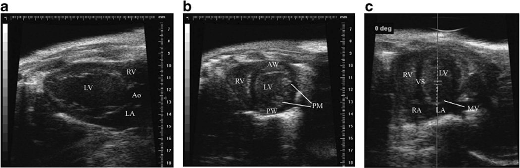
Hypertension continues to be a significant cause of morbidity and mortality, underscoring the need to better understand its early effects on the myocardium. The aim of this study is to determine the feasibility of in vivo longitudinal assessment of cardiac function, particularly diastolic function, in a mouse model of renovascular hypertension. Renovascular hypertension (RVH) was induced in 129S1/SvImJ male mice (n=9). To assess left ventricular (LV) systolic and diastolic function, M-mode echocardiography, pulsed-wave Doppler echocardiography and tissue Doppler imaging were performed at baseline, 2 and 4 weeks after the induction of renal artery stenosis. Myocardial tissue was collected to assess cellular morphology, fibrosis, extracellular matrix remodeling and inflammation ex vivo. RVH led to a significant increase in systolic blood pressure after 2 and 4 weeks (baseline: 99.26±1.09 mm Hg; 2 weeks: 140.90±7.64 mm Hg; 4 weeks: 147.52±5.91 mm Hg, P<0.05), resulting in a significant decrease in LV end-diastolic volume, associated with a significant elevation in ejection fraction and preserved cardiac output. Furthermore, the animals developed an abnormal diastolic function profile, with a shortening in the E velocity deceleration time as well as increases in the E/e' and the E/A ratio. The ex vivo analysis revealed a significant increase in myocyte size and deposition of extracellular matrix. Non-invasive high-resolution ultrasonography allowed assessment of the diastolic function profile in a small animal model of renovascular hypertension.
Figures


References
-
- Roger VL, Go AS, Lloyd-Jones DM, Benjamin EJ, Berry JD, Borden WB, Bravata DM, Dai S, Ford ES, Fox CS, Fullerton HJ, Gillespie C, Hailpern SM, Heit JA, Howard VJ, Kissela BM, Kittner SJ, Lackland DT, Lichtman JH, Lisabeth LD, Makuc DM, Marcus GM, Marelli A, Matchar DB, Moy CS, Mozaffarian D, Mussolino ME, Nichol G, Paynter NP, Soliman EZ, Sorlie PD, Sotoodehnia N, Turan TN, Virani SS, Wong ND, Woo D. Turner MBAmerican Heart Association Statistics C, Stroke Statistics S. Heart disease and stroke statistics—2012 update: a report from the American Heart Association. Circulation. 2012;125:e2–e220. - PMC - PubMed
-
- Brilla CG. Regression of myocardial fibrosis in hypertensive heart disease: diverse effects of various antihypertensive drugs. Cardiovasc Res. 2000;46:324–331. - PubMed
-
- Brilla CG, Funck RC, Rupp H. Lisinopril-mediated regression of myocardial fibrosis in patients with hypertensive heart disease. Circulation. 2000;102:1388–1393. - PubMed
-
- Brilla CG, Janicki JS, Weber KT. Cardioreparative effects of lisinopril in rats with genetic hypertension and left ventricular hypertrophy. Circulation. 1991;83:1771–1779. - PubMed
Publication types
MeSH terms
Grants and funding
LinkOut - more resources
Full Text Sources
Other Literature Sources