. 2013 Nov;84(5):920-30.
doi: 10.1038/ki.2013.175. Epub 2013 May 15.

Divergent functions of the Rho GTPases Rac1 and Cdc42 in podocyte injury

Affiliations

Divergent functions of the Rho GTPases Rac1 and Cdc42 in podocyte injury

Simone M Blattner et al. Kidney Int. 2013 Nov.

Abstract

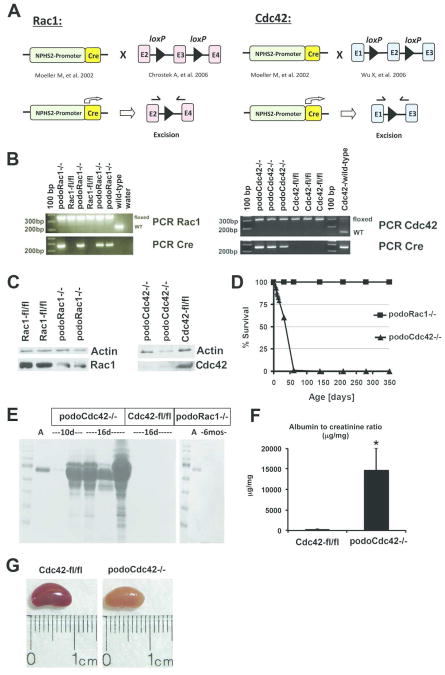
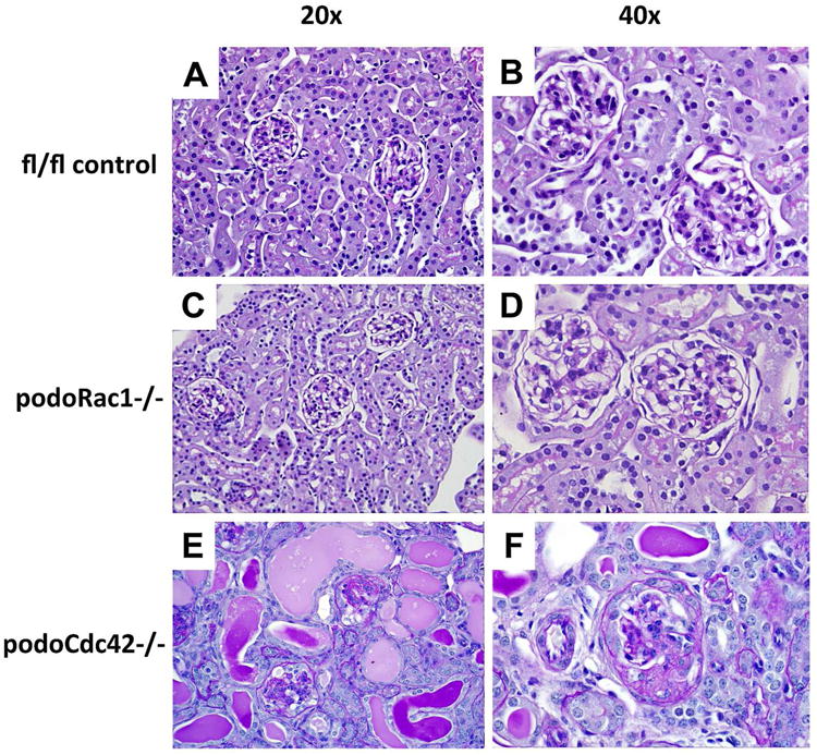
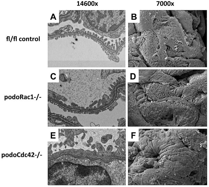
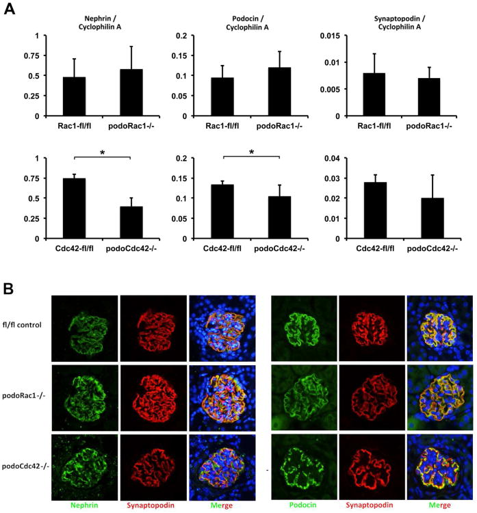
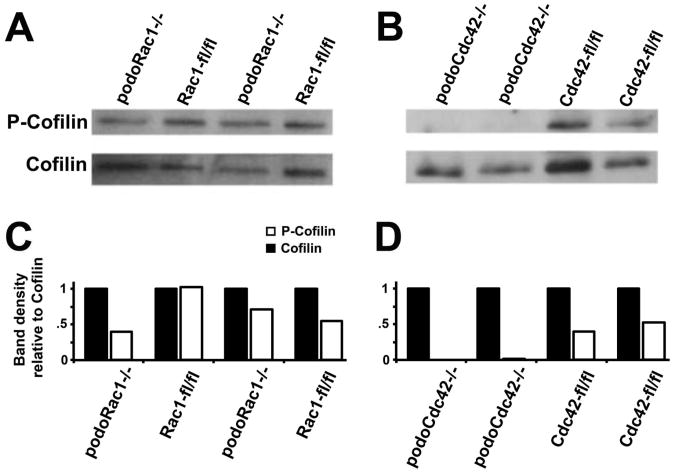
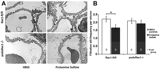
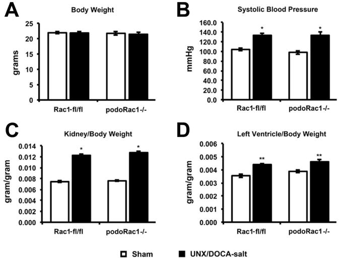
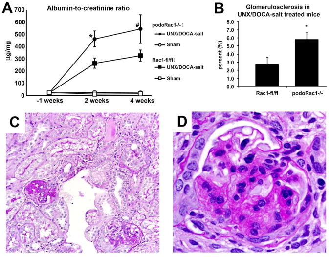
Podocytes are highly specialized epithelial cells with complex actin cytoskeletal architecture crucial for maintenance of the glomerular filtration barrier. The mammalian Rho GTPases Rac1 and Cdc42 are molecular switches that control many cellular processes, but are best known for their roles in the regulation of actin cytoskeleton dynamics. Here, we employed podocyte-specific Cre-lox technology and found that mice with deletion of Rac1 display normal podocyte morphology without glomerular dysfunction well into adulthood. Using the protamine sulfate model of acute podocyte injury, podocyte-specific deletion of Rac1 prevented foot process effacement. In a long-term model of chronic hypertensive glomerular damage, however, loss of Rac1 led to an exacerbation of albuminuria and glomerulosclerosis. In contrast, mice with podocyte-specific deletion of Cdc42 had severe proteinuria, podocyte foot process effacement, and glomerulosclerosis beginning as early as 10 days of age. In addition, slit diaphragm proteins nephrin and podocin were redistributed, and cofilin was dephosphorylated. Cdc42 is necessary for the maintenance of podocyte structure and function, but Rac1 is entirely dispensable in physiological steady state. However, Rac1 has either beneficial or deleterious effects depending on the context of podocyte impairment. Thus, our study highlights the divergent roles of Rac1 and Cdc42 function in podocyte maintenance and injury.


Figures

Figure 1
Figure 1
Targeted inactivation of Rac1 and Cdc42 in podocytes. (A) The diagram demonstrates the strategy for generation of podocyte-specific Rac1 and Cdc42 knockout mice. Mice expressing Cre-recombinase under control of the podocyte promoter (2.5P-Cre) were bred with mice carrying floxed Rac1 locus (exon 3) and floxed Cdc42 locus (exon 2). (B) PCR analysis of genomic DNA from tail clippings. The PCR product band of floxed (280 bp) and wild-type (200 bp) Rac1 as well as floxed (300 bp) and wild-type (200 bp) Cdc42 are shown. In addition, the 2.5P-Cre PCR product band (268 bp) is indicated. (C) Western blot analysis of Rac1 and Cdc42 in isolated glomeruli from podoRac1−/− and podoCdc42−/− mice with antibodies against Rac1, Cdc42, and β-actin reveals strong reduction of specific protein signal. (D) Survival curve for podoCdc42−/− and podoRac1−/− mice shows 100% mortality with loss of podocyte-specific Cdc42 by day 60. (E) SDS-PAGE analysis of urine samples demonstrates variable selective proteinuria by 10 days of age in podoCdc42−/− mice and heavy nonspecific proteinuria by 16 days of age compared to floxed control. Loss of podocyte-specific Rac1 has no effect on urine protein at 6 months. (F) Urine albumin-to-creatinine ratios in podoCdc42−/− mice were significantly increased versus floxed controls (n=5 per group). (G) Whole kidneys from podoCdc42−/− mouse at 3 weeks of age are pale yellow and display a granular surface compared to floxed control.
Figure 2
Figure 2
Progressive glomerulosclerosis in podoCdc42−/− but not podoRac1−/− mice by light microscopy (PAS staining at 20× and 40× magnification). (A, B) Floxed control mice demonstrate normal tubulointerstitial and glomerular morphology at 3-4 weeks of age. (C, D) podoRac1−/− tubulointerstitial and glomerular morphology at 12 months of age is indistinguishable from control. Podocyte-specific loss of Cdc42 results in tubular protein casts and injury (E) as well as focal segmental glomerulosclerosis in a minority of glomeruli (F) at 3-4 weeks of age.
Figure 3
Figure 3
Severe podocyte foot process effacement in podoCdc42−/− but not podoRac1−/− mice by electron microscopy (14600× transmission and 7000× scanning magnification). (A, B) Transmission and scanning electron micrographs of control mouse glomeruli show an intact arrangement of podocyte foot processes and normal filtration slits. (C, D) podoRac1−/− podocytes demonstrate no aberrant morphology and are indistinguishable from control. In contrast, podoCdc42−/− mouse podocytes show total effacement of foot processes with near absence of filtration slits. (E, F)
Figure 4
Figure 4
Glomerular mRNA and protein expression of podocyte markers from one-month-old podoRac1−/− and podoCdc42−/− mice. (A) Gene expression for podocyte markers nephrin, podocin, and synaptopodin in podoRac1−/− glomeruli does not differ from floxed control. In contrast, isolated glomeruli from podoCdc42−/− mice display a significant reduction in transcript levels of nephrin and podocin compared to floxed control mice (*p<0.05). Synaptopodin gene expression is not significantly different. (B) Immunofluorescence studies from kidney sections stained with antibodies against nephrin, podocin, and synaptopodin demonstrate continuous distribution in podocytes from floxed control and podoRac1−/− mice. In contrast, immunofluorescence for nephrin and podocin in podocytes from podoCdc42−/− mice exhibit a significantly impaired, granular distribution along the basement membrane.
Figure 5
Figure 5
Total cofilin and phospho-cofilin (p-cofilin), the active and inactive states respectively, in isolated glomeruli from podoRac1−/−, podoCdc42−/−, and floxed control mice by Western and densitometry analysis. (A and C) Compared to all floxed controls, protein expression of p-cofilin/total cofilin in glomeruli from podoRac1−/− appear similar. In stark contrast, (B and D) podoCdc42−/− glomeruli display no appreciable p-cofilin, highlighting a significant imbalance in cofilin phosphorylation.
Figure 6
Figure 6
Normal phenotype in 1-week-old podoCdc42−/− mice. (A) SDS-PAGE shows no albumin bands in urine from podocyte-specific Cdc42 knockout mice. (B) Tubulointerstitial and glomerular morphology demonstrates an immature appearance in both podoCdc42−/− and floxed control mice (PAS staining at 10× and 40× magnification). (C) Ultrastructural examination reveals no difference in glomerular capillary or podocyte foot process (inset) morphology between podoCdc42−/− and floxed control mice.
Figure 7
Figure 7
Loss of podocyte-specific Rac1 protects against induction of podocyte foot process effacement by protamine sulfate. (A) Transmission EM of glomerular capillary walls of floxed control mice after perfusion with HBSS control (top left) and protamine sulfate (top right) demonstrates partial foot process effacement (arrowheads). Glomerular capillary walls from podoRac1−/− mice after perfusion with HBSS control (bottom left) and protamine sulfate (bottom right) shows no qualitative difference in foot process morphology. Results are representative of 6-8 mice per group. ×7900 magnification (B) Filtration slit frequency per micron as seen by transmission EM reflects the morphologic interpretation. *P<0.05
Figure 8
Figure 8
Body weight, systolic blood pressure, and organ weights at 4 weeks following UNX/DOCA-salt treatment. (A) No difference in average total body weight among the four groups. (B-D) UNX/DOCA-salt treatment resulted in increased systolic blood pressure, kidney/body weight ratio, and left ventricle/body weight similarly in both podoRac1−/− and Rac1 fl/fl control mice. *P<0.01, **P<0.05
Figure 9
Figure 9
Albuminuria and percent glomerulosclerosis in UNX/DOCA-salt mice. (A) Albumin-to-creatinine ratio at 1 week before, and 2 and 4 weeks after uninephrectomy and DOCA treatment demonstrate significantly more albuminuria in podoRac1−/− mice than Rac1 fl/fl control mice. (B) UNX/DOCA-salt treated podoRac1−/− demonstrates twice the glomerulosclerosis than Rac1 fl/fl control mice. Sham treated mice of both genotypes showed no glomerulosclerosis (data not shown). (C and D) Representative PAS stained glomerulus at 20× and 40× magnification from UNX/DOCA-salt treated podoRac1−/− mouse demonstrating segmental sclerosis. *P<0.05, #P=0.19
Figure 10
Figure 10
Transmission EM of glomerular capillary walls of UNX/DOCA-salt-treated Rac1-fl/fl (A) and podoRac1−/− (B) mice displaying focal and segmental foot process effacement. Sham-treated podoRac1−/− and Rac1 fl/fl (C) exhibit only normal appearing, regularly interdigitating podocyte foot processes.

References

    1. Faul C, Asanuma K, Yanagida-Asanuma E, et al. Actin up: regulation of podocyte structure and function by components of the actin cytoskeleton. Trends Cell Biol. 2007;17:428–437. - PubMed
    1. Pavenstadt H, Kriz W, Kretzler M. Cell biology of the glomerular podocyte. Physiol Rev. 2003;83:253–307. - PubMed
    1. Heasman SJ, Ridley AJ. Mammalian Rho GTPases: new insights into their functions from in vivo studies. Nat Rev Mol Cell Biol. 2008;9:690–701. - PubMed
    1. Wennerberg K, Der CJ. Rho-family GTPases: it's not only Rac and Rho (and I like it) J Cell Sci. 2004;117:1301–1312. - PubMed
    1. Etienne-Manneville S, Hall A. Rho GTPases in cell biology. Nature. 2002;420:629–635. - PubMed

Publication types

MeSH terms