. 2013:3:1846.
doi: 10.1038/srep01846.

Snake velvet black: hierarchical micro- and nanostructure enhances dark colouration in Bitis rhinoceros

Affiliations

Snake velvet black: hierarchical micro- and nanostructure enhances dark colouration in Bitis rhinoceros

Marlene Spinner et al. Sci Rep. 2013.

Abstract

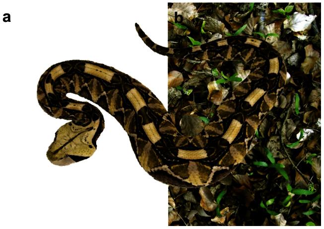
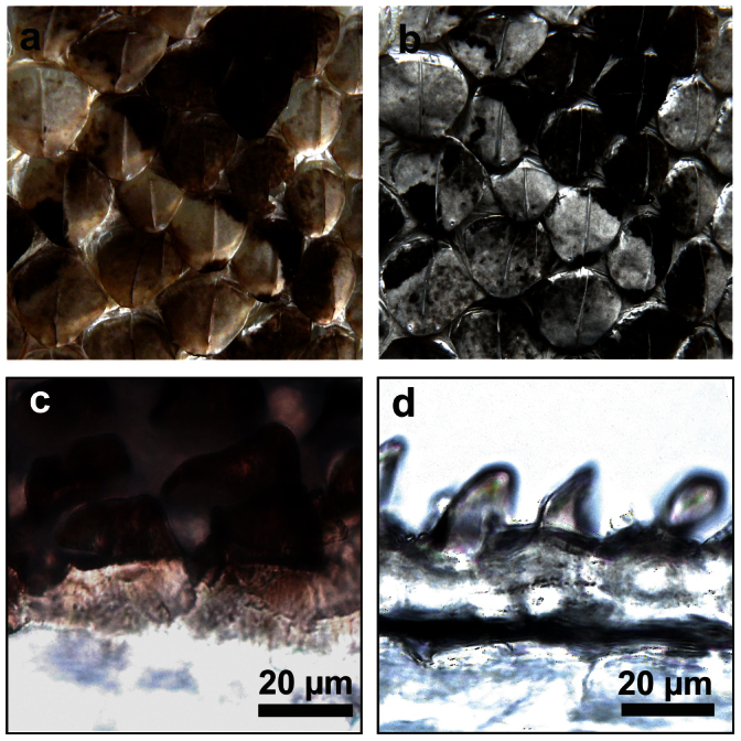
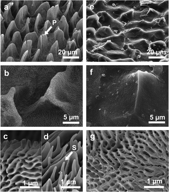
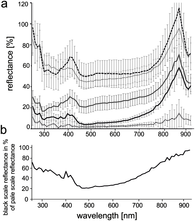
The West African Gaboon viper (Bitis rhinoceros) is a master of camouflage due to its colouration pattern. Its skin is geometrically patterned and features black spots that purport an exceptional spatial depth due to their velvety surface texture. Our study shades light on micromorphology, optical characteristics and principles behind such a velvet black appearance. We revealed a unique hierarchical pattern of leaf-like microstructures striated with nanoridges on the snake scales that coincides with the distribution of black colouration. Velvet black sites demonstrate four times lower reflectance and higher absorbance than other scales in the UV-near IR spectral range. The combination of surface structures impeding reflectance and absorbing dark pigments, deposited in the skin material, provides reflecting less than 11% of the light reflected by a polytetrafluoroethylene diffuse reflectance standard in any direction. A view-angle independent black structural colour in snakes is reported here for the first time.


Figures

Figure 1
Figure 1. Bitis rhinoceros, West African Gaboon viper (a) The snake partly on white background and (b) partly on leafy substrate similar to the natural habitat.
Figure 2
Figure 2. Colouration of scales of B. rhinoceros.
(a) Dorsal surface of exuvia of B. rhinoceros. (b) Same site sputter-coated with a 15 nm thick layer gold-palladium. (c) Light microscopy image of a cross-section from the black part of a dorsal scale of B. rhinoceros. (d) Light microscopy image of a cross-section from the pale part of a dorsal scale of B. rhinoceros.
Figure 3
Figure 3. SEM images of dorsal scales of B. rhinoceros.
(a) Microornamentation at a black dorsal scale, leaf-like structures at the surface (P). (b–c) Branched ridges at the surface of the leaf-like structures at a black dorsal scale. Ridges are regularly connected by thin struts. Pits are located between struts and ridges. (d) Ridges with spinules (S) between the leaf-like structures at a black dorsal scale. (e) Verrucate pattern at a pale dorsal scale. (f–g) Pits on the surface of a pale dorsal scale.
Figure 4
Figure 4. Reflectance of the scales of B. rhinoceros.
(a) Reflectance of black dorsal scales (black line), Au-Pd coated black dorsal scales (dotted black line), pale dorsal scales (grey line), Au-Pd coated pale dorsal scales (dotted grey line), and ventral scales (dashed black line) of B. rhinoceros in percent to the reflected light of the diffuse reflectance standard. Standard deviation of all ten scales measured is presented by error bars. (b) Reflectance of black dorsal scales in percent to the reflectance of pale scales.
Figure 5
Figure 5. Scattering measured on scales of B. rhinoceros and predicted by the Oren – Nayar model.
(a) Schematic drawing of the experimental setup for the measurement of light scattering. Intensity of scattered light was measured at different angles in respect to the scale surface normal: 0°–80° with an illumination angle at 45°. (b) Raw reflection spectra of a black (black lines) and a pale (grey lines) scale with illumination and detector arranged in the rostro-caudal direction (rc) detected from different angles. The intensity of scattered light is presented in arbitrary units. (c) Comparison between light scattering for a pale scale at 700 nm detected from lateral direction (solid line) and light scattering predicted by the model (dashed line). As fit parameters ρE0 = 37 and σ = 17.5 were applied. The light scattering intensity has the same units as in (b). (d) Intensity of light scattering for a pale scale at 700 nm detected from caudal (rc, dotted line) and lateral (l, solid line) direction. The intensity has the same units as in (b). (e) Comparison of the light scattering intensity of a pale (y-axis on the left, black line) and a black (y-axis on the right, grey line) scale at 700 nm detected from lateral direction. The intensity has the same units as in (b). (f) Light scattering angular dependence of a black scale detected from caudal (rc, dotted line) and lateral (l, solid line) direction at 700 nm. The intensity has the same units as in (b).
Figure 6
Figure 6. Transmittance of scales of B. rhinoceros.
(a) Transmittance spectra of black dorsal scales (black line), Au-Pd coated black dorsal scales (dotted black line), pale dorsal scales (grey line), Au-Pd coated pale scales dorsal (dotted grey line), and ventral scales (dashed black line) of B. rhinoceros. Standard deviation of ten scales measured is presented by error bars. (b) Transmittance of black dorsal scales (black line), Au-Pd coated black dorsal scales (dotted black line) and Au-Pd coated pale dorsal scales (dotted grey line) in percent to the pale scale transmittance.
Figure 7
Figure 7. Warm-up process of the exuvia of B. rhinoceros.
(a) Exuvia of B. rhinoceros, West African Gaboon viper. (b) IR-emission image of the exuvia of B. rhinoceros before illumination under ambient room conditions temperature, (c) after 1 min illumination, and (d) after 2 min illumination.

References

    1. Hill G. E. Female house finches prefer colourful males: sexual selection for a condition-dependent trait. Anim. Behav. 40, 563–572 (1990).
    1. Hill G. E. Female mate choice for ornamental coloration. In Bird Coloration: Vol. 2, Function and evolution (Harvard University Press, Chambridge, 2006).
    1. Milinski M. & Bakker T. C. M. Female sticklebacks use male coloration in mate choice and hence avoid parasitized males. Nature 344, 330–333 (1990).
    1. Watkins G. G. Inter-sexual signalling and the functions of female coloration in the tropidurid lizard Microlophus occipitalis. Anim. Behav. 53, 843–852 (1997).
    1. Parker A. R., Mckenzie D. R. & Ahyong S. T. A unique form of light reflector and the evolution of signalling in Ovilipes (Crustacea: Decapoda: Portunidae). Proc R Soc Lond B 265, 861–867 (1998).

Publication types

LinkOut - more resources