

Adverse local tissue reaction arising from corrosion at the femoral neck-body junction in a dual-taper stem with a cobalt-chromium modular neck
- PMID: 23677352
- PMCID: PMC3748981
- DOI: 10.2106/JBJS.L.01042
Adverse local tissue reaction arising from corrosion at the femoral neck-body junction in a dual-taper stem with a cobalt-chromium modular neck
Abstract
Background: Femoral stems with dual-taper modularity were introduced to allow additional options for hip-center restoration independent of femoral fixation in total hip arthroplasty. Despite the increasing availability and use of these femoral stems, concerns exist about potential complications arising from the modular neck-body junction.
Methods: This was a multicenter retrospective case series of twelve hips (eleven patients) with adverse local tissue reactions secondary to corrosion at the modular neck-body junction. The cohort included eight women and three men who together had an average age of 60.1 years (range, forty-three to seventy-seven years); all hips were implanted with a titanium-alloy stem and cobalt-chromium-alloy neck. Patients presented with new-onset and increasing pain at a mean of 7.9 months (range, five to thirteen months) following total hip arthroplasty. After serum metal-ion studies and metal artifact reduction sequence (MARS) magnetic resonance imaging (MRI) revealed abnormal results, the patients underwent hip revision at a mean of 15.2 months (range, ten to twenty-three months). Tissue specimens were examined by a single histopathologist, and the retrieved implants were studied with use of light and scanning electron microscopy.
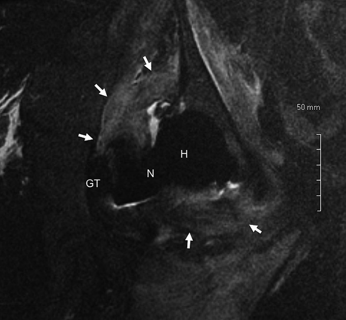
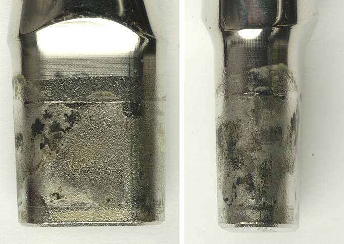
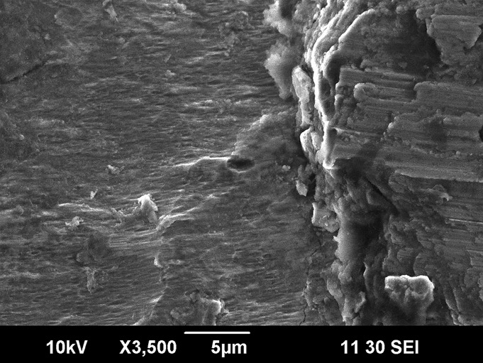
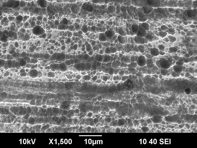
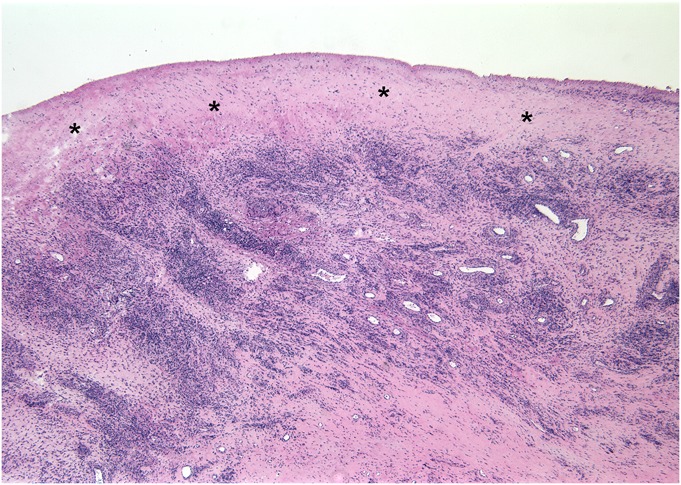
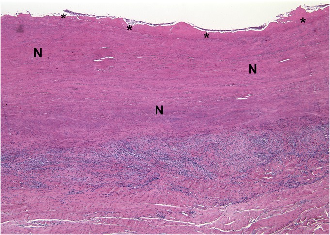
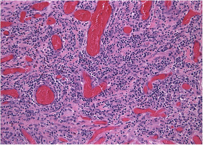
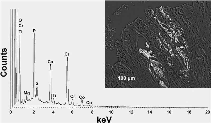
Results: Serum metal levels demonstrated greater elevation of cobalt (mean, 6.0 ng/mL) than chromium (mean, 0.6 ng/mL) or titanium (mean, 3.4 ng/mL). MRI with use of MARS demonstrated adverse tissue reactions in eight of nine patients in which it was performed. All hips showed large soft-tissue masses and surrounding tissue damage with visible corrosion at the modular femoral neck-body junction. Available histology demonstrated large areas of tissue necrosis in seven of ten cases, while remaining viable capsular tissue showed a dense lymphocytic infiltrate. Microscopic analysis was consistent with fretting and crevice corrosion at the modular neck-body interface.
Conclusions: Corrosion at the modular neck-body junction in dual-tapered stems with a modular cobalt-chromium-alloy femoral neck can lead to release of metal ions and debris resulting in local soft-tissue destruction. Adverse local tissue reaction should be considered as a potential cause for new-onset pain in patients with these components, and early revision should be considered given the potentially destructive nature of these reactions. A workup including serologic studies (erythrocyte sedimentation rate and C-reactive protein), serum metal levels, and MARS MRI can be helpful in establishing this diagnosis.
Figures
Comment in
-
That's why we call it BIOmechanics!: commentary on an article by H. John Cooper, MD, et al.: "Adverse local tissue reaction arising from corrosion at the femoral neck-body junction in a dual-taper stem with a cobalt-chromium modular neck".J Bone Joint Surg Am. 2013 May 15;95(10):e71. doi: 10.2106/JBJS.M.00169. J Bone Joint Surg Am. 2013. PMID: 23677372 No abstract available.
References
-
- Sariali E, Mouttet A, Pasquier G, Durante E, Catone Y. Accuracy of reconstruction of the hip using computerised three-dimensional pre-operative planning and a cementless modular neck. J Bone Joint Surg Br. 2009 Mar;91(3):333-40 - PubMed
-
- Duwelius PJ, Hartzband MA, Burkhart R, Carnahan C, Blair S, Wu Y, Grunkemeier GL. Clinical results of a modular neck hip system: hitting the “bull’s-eye” more accurately. Am J Orthop (Belle Mead NJ). 2010 Oct;39(10)(Suppl):2-6 - PubMed
-
- Kop AM, Swarts E. Corrosion of a hip stem with a modular neck taper junction: a retrieval study of 16 cases. J Arthroplasty. 2009 Oct;24(7):1019-23 - PubMed
Publication types
MeSH terms
Substances
LinkOut - more resources
Full Text Sources
Other Literature Sources
Medical
Research Materials
Miscellaneous
