. 2013 Jun 15;190(12):6093-103.
doi: 10.4049/jimmunol.1203048. Epub 2013 May 15.

Glia maturation factor-γ negatively modulates TLR4 signaling by facilitating TLR4 endocytic trafficking in macrophages

Affiliations

Glia maturation factor-γ negatively modulates TLR4 signaling by facilitating TLR4 endocytic trafficking in macrophages

Wulin Aerbajinai et al. J Immunol. .

Abstract

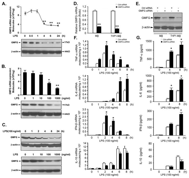
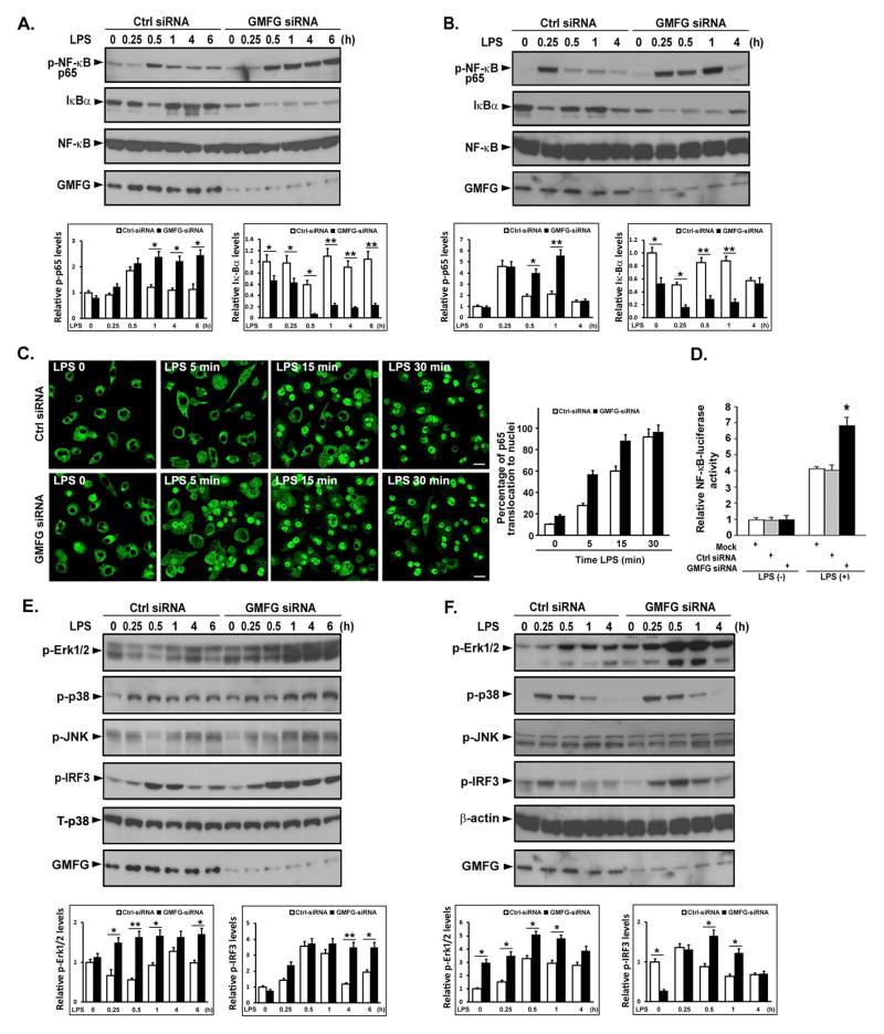
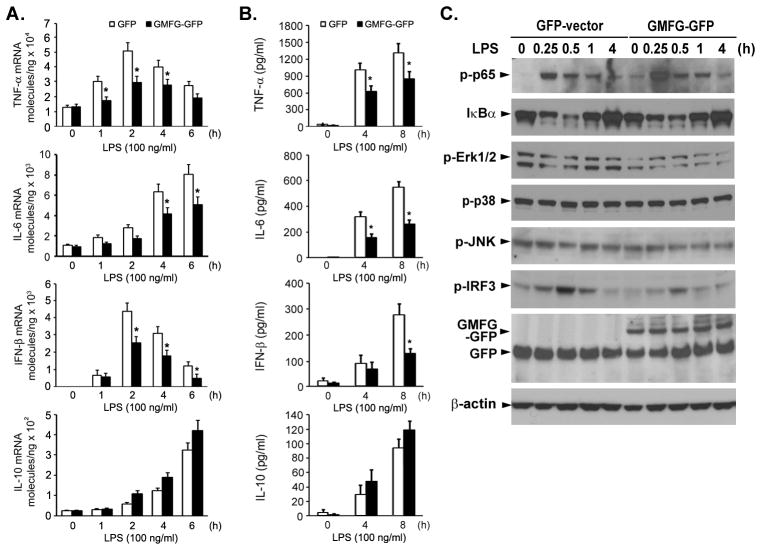
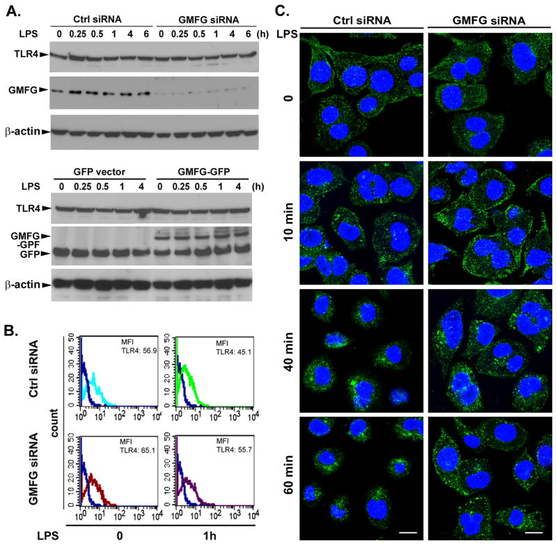
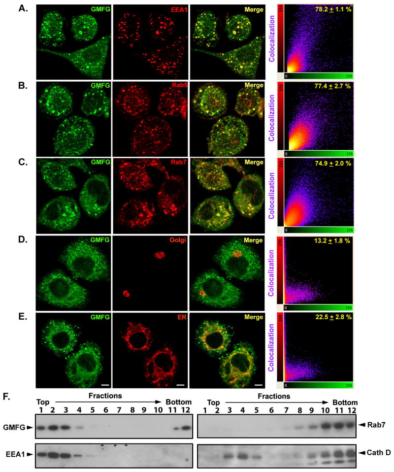
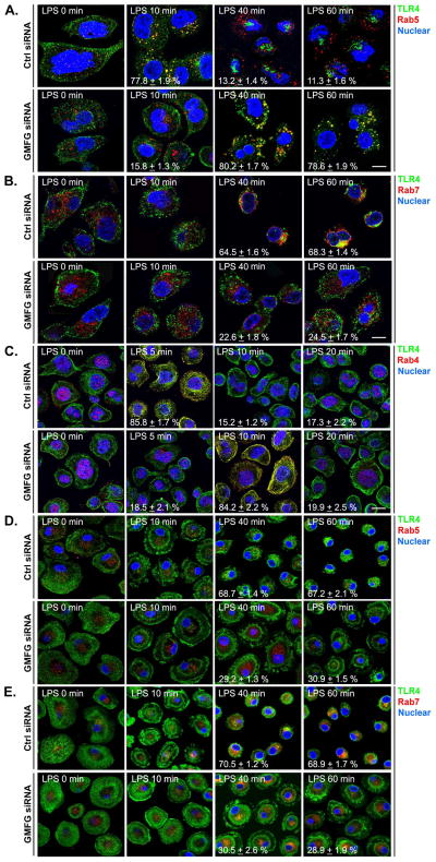
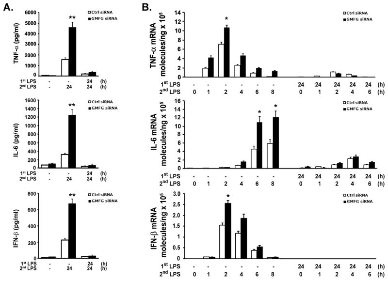
TLR4 signaling must be tightly regulated to provide both effective immune protection and avoid inflammation-induced pathology. Thus, the mechanisms that negatively regulate the TLR4-triggered inflammatory response are of particular importance. Glia maturation factor-γ (GMFG), a novel actin depolymerization factor/cofilin superfamily protein that is expressed in inflammatory cells, has been implicated in mediating neutrophil and T cell migration, but its function in macrophage immune response remains unclear. In the current study, the role of GMFG in the LPS-induced TLR4-signaling pathway was investigated in THP-1 macrophages and human primary macrophages. LPS stimulation of macrophages decreased GMFG mRNA and protein expression. We show that GMFG negatively regulates LPS-induced activation of NF-κB-, MAPK-, and IRF3-signaling pathways and subsequent production of proinflammatory cytokines and type I IFN in human macrophages. We found that endogenous GMFG localized within early and late endosomes. GMFG knockdown delayed LPS-induced TLR4 internalization and caused prolonged TLR4 retention at the early endosome, suggesting that TLR4 transport from early to late endosomes is interrupted, which may contribute to enhanced LPS-induced TLR4 signaling. Taken together, our findings suggest that GMFG functions as a negative regulator of TLR4 signaling by facilitating TLR4 endocytic trafficking in macrophages.


Conflict of interest statement

Disclosures

The authors have no conflicting financial interests.

Figures

Figure 1
Figure 1. Silencing of GMFG expression enhances LPS-induced production of proinflammatory mediators in macrophages
(A–C) GMFG expression upon LPS stimulation. Primary human macrophages (A and B) and THP-1 macrophages (C) were treated with 100 ng/ml LPS for the indicated time periods or with the indicated doses of LPS for 24 h; GMFG mRNA expression levels were assessed by Q-PCR and protein levels were determined by immunoblotting. β-actin was used as an internal control. (D and E) Primary human macrophages and THP-1 macrophages were transfected with control negative siRNA (Ctrl siRNA) or GMFG siRNA for 48 h. The efficiency of knockdown was evaluated by Q-PCR and immunoblotting analysis. (F and G) Primary human macrophages transfected with Ctrl siRNA or GMFG siRNA were treated with 100 ng/ml LPS for the indicated time periods. TNF-α, IL-6, IFN-β, and IL-10 mRNA expression levels were assessed by Q-PCR and cytokine production was measured by ELISA. Data are presented as means ± SD of three independent experiments. * p < 0.05, ** p < 0.01.
Figure 2
Figure 2. Silencing of GMFG expression potentiates the LPS-induced signaling pathway in macrophages
THP-1 macrophages and primary human macrophages were transfected with control negative siRNA (Ctrl siRNA) or GMFG siRNA. After 48 h, the cells were treated with 100 ng/ml LPS for the indicated time periods. (A and B) Cell lysates extracted from THP-1 macrophages (A) or primary human macrophages (B) were prepared and subjected to immunoblotting with the indicated Abs. NF-κB was used as an internal control. Relative quantification of phosphorylation of p65 and degradation of IκBα normalized to total NF-κB is shown in the lower panels. Data are presented as means ± SD of three independent experiments. Values for control negative siRNA (Ctrl siRNA)-transfected cells not stimulated with LPS were set to 1. * p < 0.05, ** p < 0.01. (C) Confocal-microscopy analysis of nuclear translocation of p65 after LPS stimulation in GMFG siRNA- or control negative siRNA (Ctrl siRNA)-transfected THP-1 macrophages. Scale bar, 50 μm. Quantification of p65 translocation to the nuclei is shown in the right panel. Data are presented as means ± SD of three coverslips from a representative experiment. A total of 100 cells were counted per coverslip. (D) THP-1 macrophages that had been knocked down with GMFG siRNA or control negative siRNA were transfected with pTK-Renilla-luciferase and NF-κB luciferase reporter plasmids. After 24 h, the cells were stimulated with 100 ng/ml LPS for 6 h and NF-κB luciferase activity was measured using the Dual-luciferase Reporter Assay System (Promega), normalized to the internal control renilla luciferase activity. Values for mock-transfected cells not stimulated with LPS were set to 1. Data are presented as means ± SD of three experiments. * p < 0.05. (E and F) Cell lysates extracted from GMFG siRNA- or control negative siRNA (Ctrl siRNA)-transfected THP-1 macrophages (E) or primary human macrophages (F) were subjected to immunoblotting with the indicated Abs. Total p38 and β-actin were used as internal controls. Relative quantification of phosphorylation of Erk1/2 and IκBα is shown in the lower panels. Data are presented as means ± SD of three independent experiments. Values for control negative siRNA (Ctrl siRNA)-transfected cells not stimulated with LPS were set to 1. * p < 0.05, ** p < 0.01.
Figure 3
Figure 3. Overexpression of GMFG inhibits LPS-induced cytokine production and LPS-initiated signaling pathways in macrophages
THP-1 macrophages were transfected with GMFG-GFP plasmid or GFP empty vector. After 24 h, the cells were stimulated with 100 ng/ml LPS for the indicated time periods. (A and B) TNF-α, IL-6, IFN-β, and IL-10 mRNA expression levels were measured by Q-PCR and cytokine production was measured by ELISA. Data are presented as means ± SD of three independent experiments. *, p < 0.05 compared with control cells. (C) Cell lysates from GMFG-GFP-transfected and GFP-vector-transfected THP-1 macrophages were prepared and subjected to immunoblotting with the indicated Abs. β-actin was used as an internal control.
Figure 4
Figure 4. Silencing of GMFG inhibits TLR4 internalization
(A) Immunoblotting analysis of TLR4 expression in GMFG-silenced or GMFG-overexpressed THP-1 macrophages. Cells were transfected with GMFG siRNA or control negative siRNA (Ctrl siRNA) or GFP empty vector or GMFG-GFP plasmids. After 48 h, the cells were treated with 100 ng/ml LPS for the indicated time periods, then cell lysates were prepared and subjected to immunoblotting with TLR4 or GMFG Ab. β-actin was used as an internal control. (B) Cell-surface TLR4 expression was evaluated by flow cytometry using a specific anti-TLR4 Ab in THP-1 macrophages transfected with GMFG siRNA or control negative siRNA (Ctrl siRNA). Mean fluorescence intensity is shown. (C) TLR4 localization was examined by confocal microscopy after LPS stimulation in GMFG siRNA- or Ctrl siRNA-transfected THP-1 macrophages. Cells were immunostained with anti-TLR4 Ab followed by Alexa Fluor 488-conjugated secondary Ab. Images are representative of three independent experiments. Scale bar, 100 μm.
Figure 5
Figure 5. GMFG colocalizes with multiple endosome compartments
(A–E) Confocal-microscopy analysis of THP-1 macrophages immunostained with anti-GMFG Ab (green) along with Abs for the indicated organelle membrane markers (red). Scale bar, 100 μm. Right, dual-color pixel analysis of the colocalization of GMFG and various organelle markers. Data are presented as means ± SD of three independent experiments. (F) Steady-state distribution of organelle marker proteins following subcellular fractionation using Percoll-density gradient centrifugation. Postnuclear supernatants of THP-1 macrophages were fractionated on 17% Percoll-density gradients and the individual fractions examined by immunoblotting with specific early endosome (EEA1), late endosome (Rab7), and lysosome (cathepsin D, Cell Signaling Technology) marker Abs, as well as GMFG Ab.
Figure 6
Figure 6. Silencing of GMFG induces abnormal TLR4 endosomal trafficking
THP-1 macrophages and primary human macrophages were transfected with GMFG siRNA or control negative siRNA (Ctrl siRNA). After 48 h, the cells were stimulated with or without LPS for the indicated time periods. Following treatment, cells were fixed first with 4% paraformaldehyde for 20 min, then in ice-cold methanol for another 20 min. (A–C) Fixed THP-1 macrophages were immunostained with Abs against TLR4 (green) and Rab5 (early endosome marker; red) (A), Abs against TLR4 (green) and Rab7 (late endosome marker; red) (B), or Abs against TLR4 (green) and Rab4 (fast recycling endosome marker; red) (C). (D and E) Fixed primary human macrophages were immunostained with Abs against TLR4 (green) and Rab5 (red) (D) or Abs against TLR4 (green) and Rab7 (red) (E). Nuclear DNA was labeled with DAPI (blue). Scale bar, 100μm.
Figure 7
Figure 7. GMFG is not required for endotoxin tolerance
THP-1 macrophages were transfected with GMFG siRNA or control negative siRNA (Ctrl siRNA). After 48 h, the cells were treated with LPS (1st LPS; 10 ng/ml) for 24 h, then the supernatants were removed and the cells were washed and challenged with LPS (2nd LPS; 100 ng/ml) for another 24 h. (A) TNF-α, IL-6, and IFN-β levels were measured by ELISA. (B) TNF-α, IL-6, and IFN-β mRNA expression levels were assessed by Q-PCR. Data are presented as means ± SD of three independent experiments. * p < 0.05, ** p < 0.01.

References

    1. Akira S, Uematsu S, Takeuchi O. Pathogen recognition and innate immunity. Cell. 2006;124:783–801. - PubMed
    1. West AP, Koblansky AA, Ghosh S. Recognition and signaling by toll-like receptors. Annu Rev Cell Dev Biol. 2006;22:409–437. - PubMed
    1. Kawai T, Akira S. The role of pattern-recognition receptors in innate immunity: update on Toll-like receptors. Nat Immunol. 2010;11:373–384. - PubMed
    1. Akira S, Takeda K. Toll-like receptor signalling. Nat Rev Immunol. 2004;4:499–511. - PubMed
    1. Barrat FJ, Coffman RL. Development of TLR inhibitors for the treatment of autoimmune diseases. Immunol Rev. 2008;223:271–283. - PubMed

Publication types