. 2014 Mar;66(2):283-97.
doi: 10.1007/s10616-013-9568-0. Epub 2013 May 16.

Assessment of anti-mutagenic, anti-histopathologic and antioxidant capacities of Egyptian bee pollen and propolis extracts

Affiliations

Assessment of anti-mutagenic, anti-histopathologic and antioxidant capacities of Egyptian bee pollen and propolis extracts

Amany A Tohamy et al. Cytotechnology. 2014 Mar.

Abstract

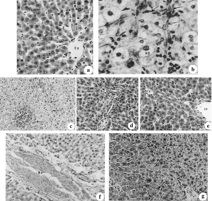
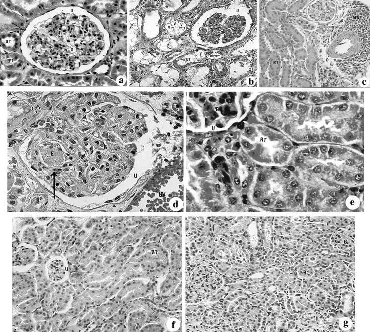
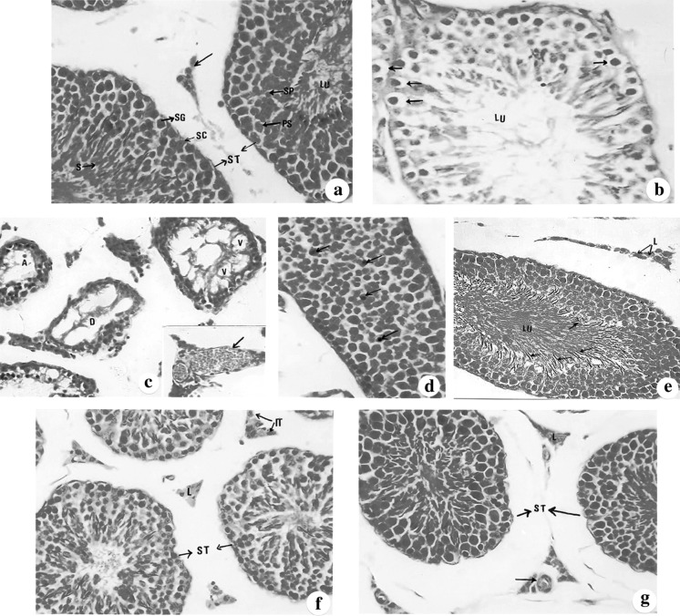
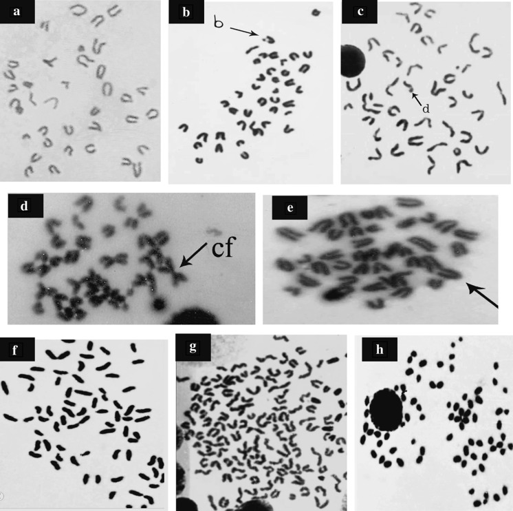
Bee pollen and propolis are popular, traditional health foods. The objective of the current study was to investigate the anti-mutagenic, anti-histopathologic and antioxidant effects among water extracts of Egyptian bee pollen (WEBP) and brown powder of water-soluble derivative propolis (WSDP) on cisplatin (CDDP) induced hepatic, renal, testicular and genotoxicity in male albino mice (Mus muscullus), in addition to their effects on the oxidant/antioxidant status in the tested organs. Hepatic, renal and testicular dysfunctions were evaluated histologically; while genotoxicity and cytotoxicity were evaluated by the bone marrow chromosomal aberration assay and mitotic index, respectively. Moreover, oxidative stress was explored via determination of lipid peroxidation, catalase activity and the concentration of the reduced form of glutathione. The treatment of mice with WEBP and WSDP at doses 140 and 8.4 mg/kg b. wt./day, respectively for 14 days simultaneously with CDDP (2.8 mg/kg b. wt.) resulted in significant protection. The positive control animals taken CDDP alone showed toxic histological and genetical manifestations (at P < 0.05) accompanied with an elevated content of peroxidized lipid and lowered catalase activity and glutathione concentration in the homogenate of liver, kidney and testis tissues (at P < 0.001). These toxic side effects in all tested organs were greatly ablated with a significant reduction in lipid peroxidation level and elevation in catalase activity and glutathione concentration (P < 0.001) when using both WEBP and WSDP. On the basis of the present assays, Bee pollen appears more potent in exerting an ameliorative effect and this effect was more pronounced in testis.


Figures

Fig. 1
Fig. 1
(a–g): Photomicrographs of liver sections of mice stained with haematoxylin and eosin showing a control liver section showing the central vein (CV), the hepatic sinusoids (S), the hepatic strands (H) and the Kupffer cells (X 128), b hydrobic degenerative changes (HD), fibroblastic proliferation (downward arrow) seen in the hepatic parenchyma of cisplatin injected mice for twenty-one days (×512), c necrotic foci (N) seen in the parenchyma with hydrobic degenerative changes (downward arrow) of liver section of cisplatin-treated mice surrounded by highly eosinophilic hepatocytes and occupied with mononuclear leucocytes (×100), d minimal fibrosis (downward arrow) seen in the portal area of propolis-treated animals (X 128), e normal hepatic tissue organization registered in bee pollen-treated mice (×128), f periportal odema (O) and portal vein congestion with normal hepatocytes organization and appearance, recorded in animals treated with propolis after cisplatin administration (×100) and g diffuse proliferation of Kupffer cells (downward arrow) noticed in the hepatic parenchyma in the central area of animals treated with bee pollen after cisplatin injection (×128)
Fig. 2
Fig. 2
(a–g): Showed sections of kidney in different examined groups stained with haematoxylin and eosin. a A photomicrograph of the cortical region of a kidney of control mice showing the golmerulus (G) with its Bowman’s capsule (downward arrow), urinary space (U), glomerular apparatus (GA), the proximal tubule (PT) and distal tubule (DT) (×400). b A photomicrograph of the cortical region of kidney section of mice administered cisplatin illustrating widening of the urinary space (U), atrophy of the glomerulus (G), and vacuolar degenerative changes in the renal tubules (V) (×1,000). c A light micrograph showing perivascular focal mononuclear leucocytic aggregation (MN) and odema (O). Renal tubular (RT) epithelial cells showed hyperplasia with obliterated lumen (×400). d A light micrograph of the glomerulus of propolis-treated group showing eosinophilic bodies (E) (downward arrow) and congested blood vessels (BV) (×400). e A photomicrograph of the kidney section of administered bee pollen showing nearly normal cortical structure. (G: glomerulus, U: urinary space, RT: renal tubule) (×400). f Lightmicrogarph of the kidney section of mice administered cisplatin and treated with propolis having renal tubular (RT) normal structures with hyperemic glomeruli (G) (×150). g A photomicrograph of the kidney of mice given cisplatin and treated with bee pollen illustrating normal renal tubules (RT) and glomeruli (G) (×150)
Fig. 3
Fig. 3
(a–g): Sections of testis in different examined groups stained with haematoxylin and eosin. a A control seminiferous tubules (ST) and interstitial cells (downward arrow) showing the normal spermatogenic lineage; spermatogonia (SG). Note the sertoli cells (SC) are seen between the spermatogonia. LU: lumen (×400). b A photomicrograph of the testis section of cisplatin injected mice showing the seminiferous tubule with few number of spermatogenic lineage and numerous vacuolated spermatogonia (downward arrow) LU: lumen (×1,000). c A lightmicrograph of the testis section of a cisplatin-injected mice showing atrophied (A) seminiferous tubules or degenerated ones (D) with severe vacuolation (V).The insertion: congested blood vessel in the interstitial tissue (×400). d A photomicrograph of the testis section of mice administered propolis showing a normal seminiferous tubule invaded with macrophages (downward arrow) (×400). e A light micrograph of a testis section of mice given bee pollen illustrating normal interstitial tissue with Leydig cells (L), seminiferous tubules with respect number of sperms (downward arrow).Lu: lumen (×400). f A light micrograph of the testis section of mice given cisplatin and treated with bee pollen illustrating control seminiferous tubules (ST) and interstitial tissues (IT) with Leydig cells (L) (×400). g A photomicrograph of the testis section of mice injected with cisplatin and treated with propolis showing normal seminiferous tubules (ST) with normal lineage and interstitial tissue with Leydig cells (L) and blood vessels (downward arrow) (×400)
Fig. 4
Fig. 4
(A–H): Metaphase spreads from mouse bone marrow cells showing A normal cell, B chromatid break (b), C chromatid deletion (d), D centric fusion (cf), E end to end association (arrow), F centric attenuation, G polyploidy and H endomitosis (1,000)

References

    1. Abdella E, Ahmed R. Suppression of doxorubicin apoptotic, histopathologic, mutagenic and oxidative stress effect in mice bone marrow and tested tissue by aqueous Rosemary leave extract. Egypt J Zool. 2008;51:305–330.
    1. Ahmed R, Abdella E. Modulatory effect of Rosemary leave aqueous extract on doxorubicin-induced histological lesions, apoptosis and oxidative stress in mice. J Egypt Ger Soc Zool. 2009;57C:105–137.
    1. Aly M, Ashour M, El Nahas S, Abo Zeid M. Genotoxicity and cytotoxicity of the anticancer drugs gemcitabine and cisplatin, separately and in comination: in vivo studies. J Biol Sci. 2003;3(11):961–972. doi: 10.3923/jbs.2003.961.972. - DOI
    1. Amin A, Hamza A. Effects of Roselle and Ginger on cisplatin-induced reproductive toxicity in rats. Asian J Androl. 2006;8(5):607–612. doi: 10.1111/j.1745-7262.2006.00179.x. - DOI - PubMed
    1. Awale S, Shrestha SP, Tezuka Y, Ueda J, Matsushige K, Kadota S. Neoflavonoids and related constituents from Nepalese propolis and their nitric oxide production inhibitory activity. J Nat Prod. 2005;68:858–864. doi: 10.1021/np050009k. - DOI - PubMed

LinkOut - more resources