. 2013 Jul;41(13):6501-13.
doi: 10.1093/nar/gkt397. Epub 2013 May 15.

The helicase FBH1 is tightly regulated by PCNA via CRL4(Cdt2)-mediated proteolysis in human cells

Affiliations

The helicase FBH1 is tightly regulated by PCNA via CRL4(Cdt2)-mediated proteolysis in human cells

Agathe Bacquin et al. Nucleic Acids Res. 2013 Jul.

Abstract

During replication, DNA damage can challenge replication fork progression and cell viability. Homologous Recombination (HR) and Translesion Synthesis (TLS) pathways appear as major players involved in the resumption and completion of DNA replication. How both pathways are coordinated in human cells to maintain genome stability is unclear. Numerous helicases are involved in HR regulation. Among them, the helicase FBH1 accumulates at sites of DNA damage and potentially constrains HR via its anti-recombinase activity. However, little is known about its regulation in vivo. Here, we report a mechanism that controls the degradation of FBH1 after DNA damage. Firstly, we found that the sliding clamp Proliferating Cell Nuclear Antigen (PCNA) is critical for FBH1 recruitment to replication factories or DNA damage sites. We then showed the anti-recombinase activity of FBH1 is partially dependent on its interaction with PCNA. Intriguingly, after its re-localization, FBH1 is targeted for degradation by the Cullin-ring ligase 4-Cdt2 (CRL4(Cdt2))-PCNA pathway via a PCNA-interacting peptide (PIP) degron. Importantly, expression of non-degradable FBH1 mutant impairs the recruitment of the TLS polymerase eta to chromatin in UV-irradiated cells. Thus, we propose that after DNA damage, FBH1 might be required to restrict HR and then degraded by the Cdt2-proteasome pathway to facilitate TLS pathway.


Figures
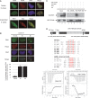
Figure 1.
Figure 1.
FBH1 interacts with PCNA via two distinct motifs, PIP-box and APIM. (A) MRC5 cells expressing ectopic FBH1 were fixed and co-stained for FBH1 (green) and PCNA (red) or EdU (red). DNA is visualized in blue. Representative images are shown for each condition. (B) MRC5 cells expressing HA-FBH1 were locally UV irradiated at 100 J/m2 and co-stained for FBH1 (green), PCNA (red) and DNA (blue) at indicated time. Representative images are shown. Each image represents >600 FBH1-positive cells in three independent experiments. The graph shows the percentage of green cells displaying HA-FBH1 accumulation to local irradiation area. Mean of three independent experiments (± SD). (C) Immunoprecipitation against HA-tag or PCNA was performed from extracts of 293T cells co-expressing HA-FBH1 and GFP-PCNA and analysed by western-blotting. Asterisk denoted aspecific signal. (D) Schematic of FBH1 protein with its PIP-box and APIM motif. Punctual mutations used for functional study are shown (top panel). Sequence alignment of the PIP-box and APIM motif of known proteins. Canonical residues are shown in red (middle panel). Thermogram and binding isotherm of titration of FBH1 PIP box wt (right) and APIM wt (left) peptides into PCNA solution were assessed by ITC at 6°C (bottom panel).
Figure 2.
Figure 2.
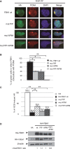
The PIP-box and APIM motif are required for FBH1 recruitment to sites of DNA damage and partially for its anti-recombinogenic activity. (A and B) MRC5 expressing HA-FBH1 wt or indicated mutants were locally irradiated at 100 J/m2 then co-stained for HA (green), PCNA (red) and DNA (blue) 3 h later. The graph shows the percentage of green cells in which FBH1 co-localizes with PCNA. Mean of three independent experiments (±SD), *P < 0.05 and **P < 0.005 by Student’s t-test. (C and D) The effects of exogenous HA-FBH1wt and indicated mutants on recombination induced by I-SceI were measured in RG37 cells as the amount of GFP-positive cells by flow cytometry analysis, in comparison with control cells expressing the empty vector. The values correspond to the mean percentage of HR efficiency, the control condition being arbitrary fixed to 100%. Mean of four independent experiments (±SD), **P < 0.005 by Student’s t-test. In parallel, the levels of exogenous FBH1 and I-Sce1 were estimated by western blot using anti-HA antibody.
Figure 3.
Figure 3.
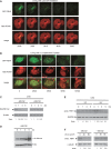
FBH1 is unstable at DNA replication sites or upon UV irradiation. (A) MRC5 cells stably expressing Halo-PCNA were transfected with GFP-FBH1. After staining of Halo-PCNA with TMR ligand, cells were locally irradiated then live cells imaged 1.30 h later. Representative distributions of GFP-FBH1 and PCNA are shown at indicated time. (B) As in (A) in non-irradiated S-phase cells. (C) MRC5 cells expressing HA-FBH1 were treated with cycloheximide (CHX) with or without MG132. Protein levels of HA-FBH1 were monitored by immunoblotting with anti-HA antibody. β-actin shows equal protein loading. Ratio of HA-FBH1 to β-actin levels is presented for each time-point with value arbitrary fixed to 1 for time 0 h. (D) Slower migrating bands detected by immunoblotting with FBH1 antibody from cells expressing HA-FBH1wt and treated with MG132 for indicated time. (E) HeLa cells were irradiated or not at 50 J/m2 then incubated with CHX. Protein levels of FBH1 and GAPDH were monitored with specific antibodies. (F) HeLa cells were irradiated with UV-C doses ranging from 0 to 50 J/m2 and subsequently incubated or not in MG132. Levels of FBH1 and γ-tubulin were monitored with specific antibodies. Ratio of FBH1 to γ-tubulin levels is presented. The experiments displayed in (C), (E) and (F) were performed at least three times, and each blot shows representative result.
Figure 4.
Figure 4.
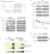
FBH1 is targeted by CRL4-Cdt2 and PCNA for degradation via a non-canonical PIP degron. (A) Alignment of PIP degron containing proteins and putative PIP degron of FBH1. Canonical PIP residues (red), ‘Degron-specific’ basic residue at +4 (green) and ‘TD motif’ (blue) are figured, with mutant sequences used in this study. (B) MRC5 cells stably expressing HA-FBH1 wt or a PIP degron mutant (PIPdeg6A) were transfected with non-targeted siRNA (NT) or siRNAs targeting Cdt2, DDB1, Cul4A and B, or PCNA. Levels of HA-FBH1, Cdt1 and proteins depleted by siRNAs were monitored with specific antibodies. (C) 293T cells were transiently co-transfected with wt or a PIP degron mutant (PIPdeg3A) of FBH1, and His-tagged ubiquitin when indicated. Cells were then incubated or not with MG132, and His-ubiquitinated (His-Ub) proteins were Ni2+ pulled-down in denaturing conditions. His-Ub FBH1 levels were immunodetected by western blotting with anti-FBH1 antibody. (D) MRC5 cells stably expressing HA-FBH1 wt were transfected with non-targeted siRNA (NT) or siRNAs targeting Cdt2. Seventy-two hours later, cells were irradiated or not at 50 J/m2 and harvested at indicated time. Levels of HA-FBH1, Cdt1 and Cdt2 were monitored with specific antibodies. The blots were quantified using an image reader. (E) MRC5 cells transfected with HA-FBH1 wt or HA-FBH1-PIPdegK+4A were irradiated at 50 J/m2, and FBH1 levels were detected from chromatin fraction (Chr.) at indicated time using HA antibody. Lamin A shows equal protein loading. The experiments displayed in (D) and (E) were carried out at least three times, and each blot shows representative result.
Figure 5.
Figure 5.
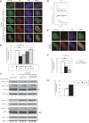
Forced expression of FBH1 or failure to degrade FBH1 via CRL4-Cdt2–PCNA pathway impairs Polη recruitment on chromatin upon UV irradiation. (A) MRC5 cells transfected with GFP-polη and indicated HA-FBH1 constructs were UV irradiated at 20 J/m2. Then 3 h later, the subnuclear localisation of GFP-polη (green) and HA-FBH1 (red) was analysed by immunofluorescence. Representative images are shown. (B) The graph shows the percentage of transfected cells displaying Polη foci from experiment described in (A). Mean of three independent experiments (±SD), *P < 0.05 by Student’s t-test. (C) MRC5 cells transfected with GFP-polη and either HA, HA-FBH1wt or HA-FBH1 PIP+APIM plasmids, were mock-treated or UV irradiated at 20 J/m2. Three and six hours later, whole cell extracts (WCE) and insoluble fractions (Chr.) were collected and analysed with indicated antibodies. (D) Thermogram and binding isotherm of titration of FBH1 PIP degK+4A peptide into PCNA solution was assessed by ITC at 6°C as described in ‘Materials and Methods’ section. (E) As in (A) with indicated constructs. Representative images are shown. (F) The graph shows the percentage of transfected cells displaying Polη foci from experiment described in (E). Mean of three independent experiments (±SD) *P < 0.05 by Student’s t-test. (G) MRC5 cells transfected with siRNA (unspecific NT or against FBH1) and GFP-polη were UV irradiated at 20 J/m2. Then 3 h later, the subnuclear localisation of GFP-polη (green) and PCNA (red) was analysed by immunofluorescence. The graph shows the percentage of transfected cells displaying Polη foci. Mean of three independent experiments (±SD).

References

    1. Li X, Heyer WD. Homologous recombination in DNA repair and DNA damage tolerance. Cell Res. 2008;18:99–113. - PMC - PubMed
    1. Branzei D, Foiani M. RecQ helicases queuing with Srs2 to disrupt Rad51 filaments and suppress recombination. Genes Dev. 2007;21:3019–3026. - PubMed
    1. Krejci L, Van Komen S, Li Y, Villemain J, Reddy MS, Klein H, Ellenberger T, Sung P. DNA helicase Srs2 disrupts the Rad51 presynaptic filament. Nature. 2003;423:305–309. - PubMed
    1. Veaute X, Jeusset J, Soustelle C, Kowalczykowski SC, Le Cam E, Fabre F. The Srs2 helicase prevents recombination by disrupting Rad51 nucleoprotein filaments. Nature. 2003;423:309–312. - PubMed
    1. Chiolo I, Saponaro M, Baryshnikova A, Kim JH, Seo YS, Liberi G. The human F-Box DNA helicase FBH1 faces Saccharomyces cerevisiae Srs2 and postreplication repair pathway roles. Mol. Cell. Biol. 2007;27:7439–7450. - PMC - PubMed

Publication types

MeSH terms