. 2013 Jun;2(6):464-72.
doi: 10.5966/sctm.2012-0144. Epub 2013 May 15.

Neural precursor cells cultured at physiologically relevant oxygen tensions have a survival advantage following transplantation

Affiliations

Neural precursor cells cultured at physiologically relevant oxygen tensions have a survival advantage following transplantation

Sybil R L Stacpoole et al. Stem Cells Transl Med. 2013 Jun.

Abstract

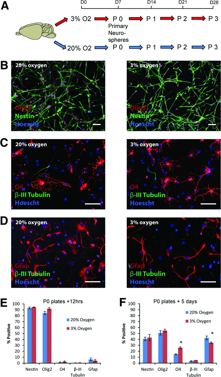
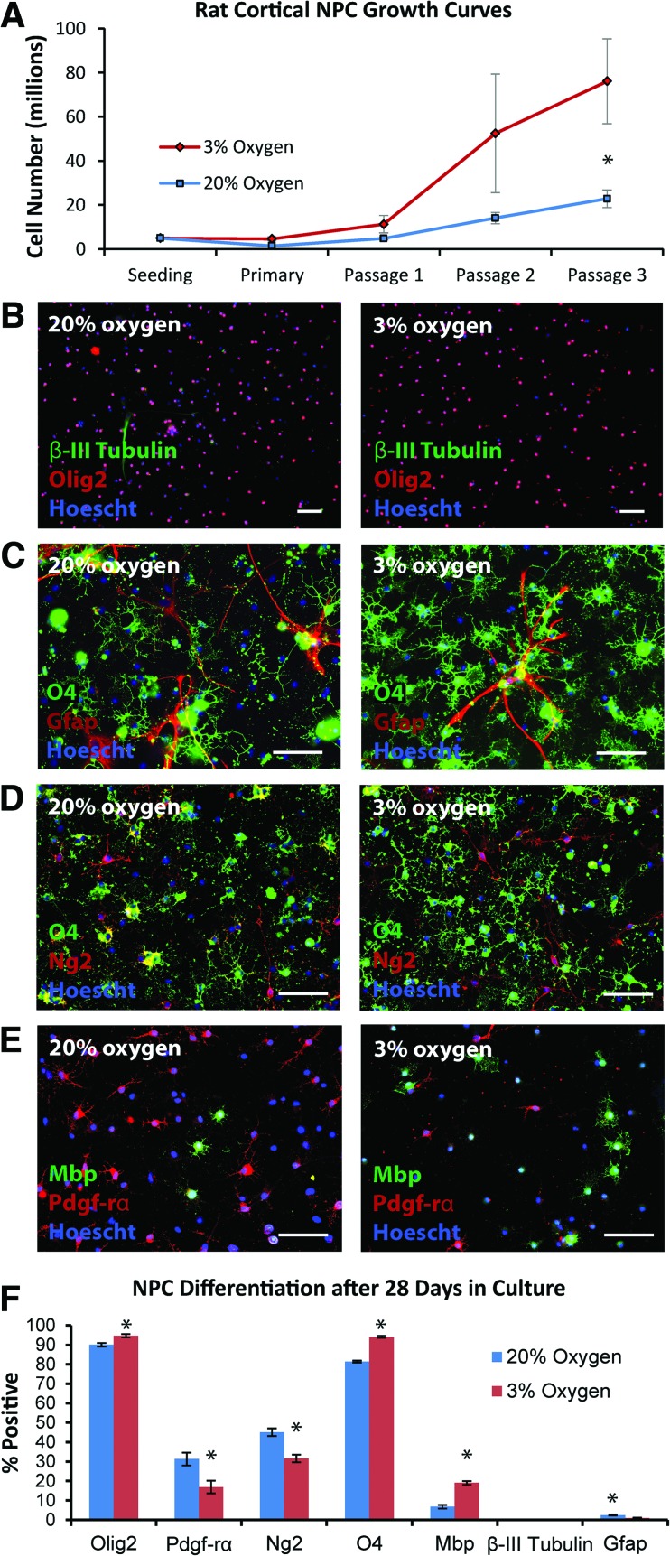
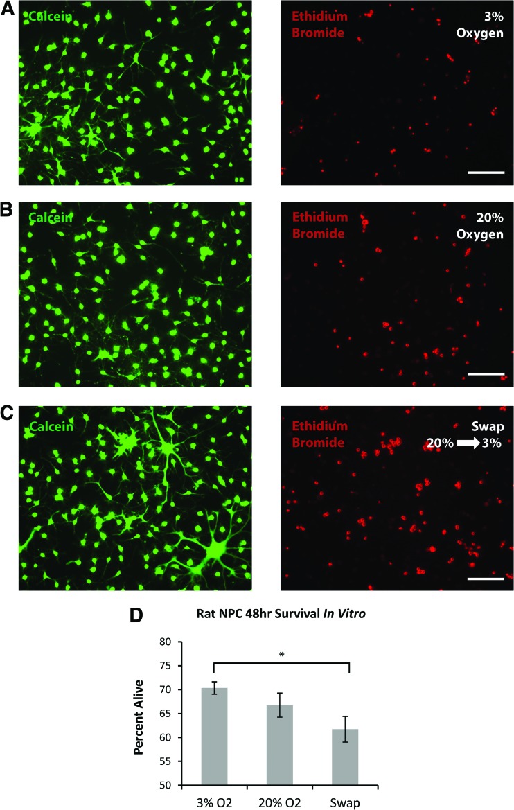
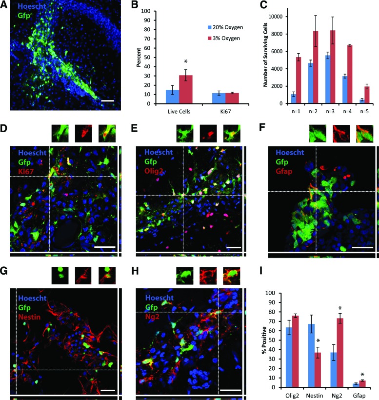
Traditionally, in vitro stem cell systems have used oxygen tensions that are far removed from the in vivo situation. This is particularly true for the central nervous system, where oxygen (O2) levels range from 8% at the pia to 0.5% in the midbrain, whereas cells are usually cultured in a 20% O2 environment. Cell transplantation strategies therefore typically introduce a stress challenge at the time of transplantation as the cells are switched from 20% to 3% O2 (the average in adult organs). We have modeled the oxygen stress that occurs during transplantation, demonstrating that in vitro transfer of neonatal rat cortical neural precursor cells (NPCs) from a 20% to a 3% O2 environment results in significant cell death, whereas maintenance at 3% O2 is protective. This survival benefit translates to the in vivo environment, where culture of NPCs at 3% rather than 20% O2 approximately doubles survival in the immediate post-transplantation phase. Furthermore, NPC fate is affected by culture at low, physiological O2 tensions (3%), with particularly marked effects on the oligodendrocyte lineage, both in vitro and in vivo. We propose that careful consideration of physiological oxygen environments, and particularly changes in oxygen tension, has relevance for the practical approaches to cellular therapies.

Keywords: Cell transplantation; Hypoxia; Neural stem cell; Oligodendrocytes.


Figures

Figure 1.
Figure 1.
Characterization of rat cortical neural precursor cell (NPC) cultures established at 3% versus 20% oxygen. (A): Primary rat neonatal cortical neurospheres were established after 7 days in culture at either 20% or 3% O2. (B, E): Following dissociation of these passage 0 primary neurospheres to single cells, the majority of NPCs labeled with nestin and olig2, with no significant difference between the two culture conditions (n = 4–5 per group). (C, D, F): After 5 days of differentiation at either 20% or 3% O2, all three neural lineages were generated, with a significant increase in O4-positive oligodendrocytes (p = .001) at 3% compared with 20% O2, along with a concomitant decrease in gfap-positive astrocytes (p = .048). Scale bars = 50 μm. Pictures shown are from NPCs derived from green fluorescence protein (GFP)-negative littermates for ease of representation (no significant differences were observed between GFP+ and GFP− NPC cultures). Abbreviations: Gfap, glial fibrillary acidic protein; P, passage.
Figure 2.
Figure 2.
Longer term expansion of rat cortical NPCs at 3% versus 20% O2 demonstrates increased generation of oligodendrocytes at 3% O2. (A): Rat cortical NPCs were expanded at either 3% or 20% O2, with greater NPC numbers observed by passage 3 at 3% O2 (p = .05; n = 3–4 per group). (B, C): NPCs plated for differentiation for 48 hours after 28 days in culture tended to generate oligodendrocyte lineage cells, rather than neurons or astrocytes, although significantly more O4-positive oligodendrocytes were generated at 3% O2. (D, E): These oligodendrocyte lineage cells matured more rapidly when expanded and differentiated at 3% versus 20% O2, as indicated through ng2, pdgf-rα, and mbp staining. (F): Quantification of day 28 NPC fate after 48 hours of differentiation; p values are .04 (olig2), .03 (pdgf-rα), .04 (ng2), <.0001 (O4), <.0001 (mbp), .45 (β-III tubulin), and .003 (gfap); n = 4–8 per group. Scale bars = 100 μm (B) and 50 μm (C–E). Abbreviations: Gfap, glial fibrillary acidic protein; Mbp, myelin basic protein; NPC, neural precursor cell; Pdgf-rα, platelet-derived growth factor receptor-α.
Figure 3.
Figure 3.
In vitro modeling of the oxygen switch that occurs during transplantation shows that switching neural precursor cells (NPCs) from 20% to 3% O2 causes significant cell death. (A–C): Representative imaging of calcein and ethidium bromide labeling of the same field of view, after 48 hours of differentiation of day 23 NPCs at 3% O2, at 20% O2, or swapped from 20% to 3% O2. (D): Quantification demonstrated a significant increase in cell death in differentiating NPCs switched from 20% to 3% O2 compared with those maintained at 3% O2 throughout (p = .007; n = 4 per group). Scale bars = 50 μm.
Figure 4.
Figure 4.
Transplantation of rat cortical neural precursor cells (NPCs) into the adult rat hippocampus shows a survival advantage and increased differentiation towards the oligodendrocyte lineage for NPCs expanded at 3% compared with 20% O2. (A): A typical graft of green fluorescence protein (GFP)+ rat cortical NPCs is shown at low magnification, 48 hours after transplantation. (B): A significantly greater percentage of GFP+ cells survived from the rat cortical NPCs maintained at 3% compared with 20% O2; there was no difference in proliferation between the two groups. (C): Total cell numbers from each individual animal further demonstrate the enhanced survival seen with the NPCs expanded at 3% O2. (D–H): Representative images demonstrating colabeling between GFP and either Ki67, olig2, gfap, nestin, and ng2 are shown. All images are from animals that received NPCs cultured at 3% O2; individual colabeled cells are shown above the main images. (I): Quantification of expression of olig2, nestin, ng2, and gfap by grafted GFP+ cells derived from NPCs expanded at 3% versus 20% O2. p values are .003 (live cells), .95 (Ki67), .12 (olig2), .02 (nestin), .03 (ng2), and 0.04 (gfap). Scale bars = 50 μm (A) and 25 μm (D–H). Abbreviations: Gfap, glial fibrillary acidic protein; Gfp, green fluorescent protein.

Similar articles

Cited by

References

    1. Smith HK, Gavins FN. The potential of stem cell therapy for stroke: Is PISCES the sign? FASEB J. 2012;26:2239–2252. - PubMed
    1. Martino G, Franklin RJM, Baron Van Evercooren A, et al. Stem cell transplantation in multiple sclerosis: Current status and future prospects. Nat Rev Neurol. 2010;6:247–255. - PubMed
    1. Cova L, Ratti A, Volta M, et al. Stem cell therapy for neurodegenerative diseases: The issue of transdifferentiation. Stem Cells Dev. 2004;13:121–131. - PubMed
    1. Brundin P, Barker RA, Parmar M. Neural grafting in Parkinson's disease: Problems and possibilities. Prog Brain Res. 2010;184:265–294. - PubMed
    1. Brundin P, Karlsson J, Emgard M, et al. Improving the survival of grafted dopaminergic neurons: A review over current approaches. Cell Transplant. 2000;9:179–195. - PubMed

Publication types

MeSH terms

LinkOut - more resources