. 2013 Aug;7(3):207-17.
doi: 10.1007/s12079-013-0202-x. Epub 2013 May 16.

Regulation of CCN1 via the 3'-untranslated region

Affiliations

Regulation of CCN1 via the 3'-untranslated region

Yosuke Nakagawa et al. J Cell Commun Signal. 2013 Aug.

Abstract

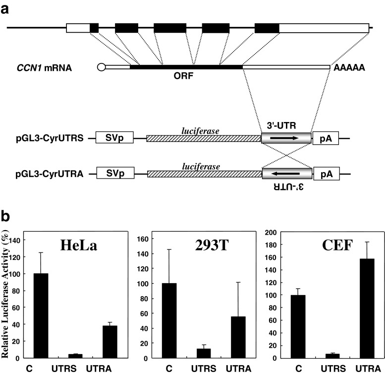
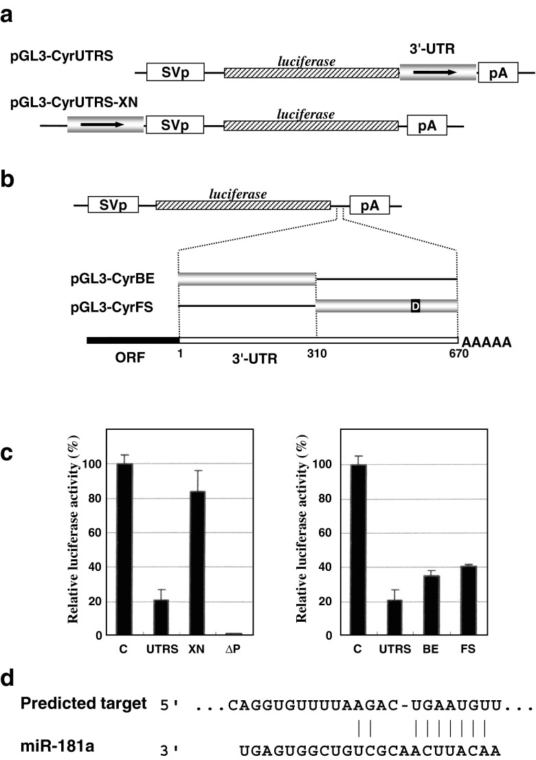
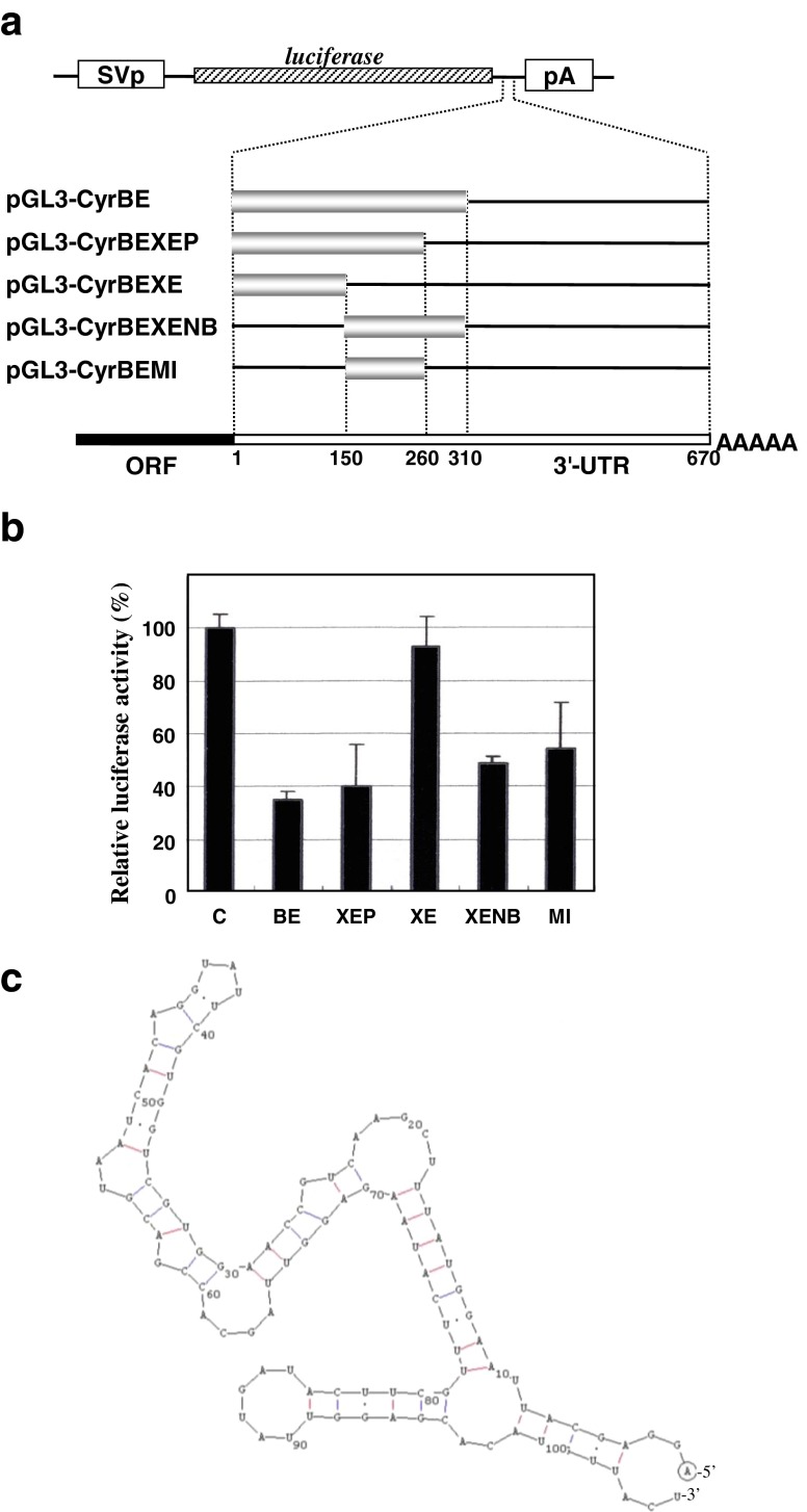
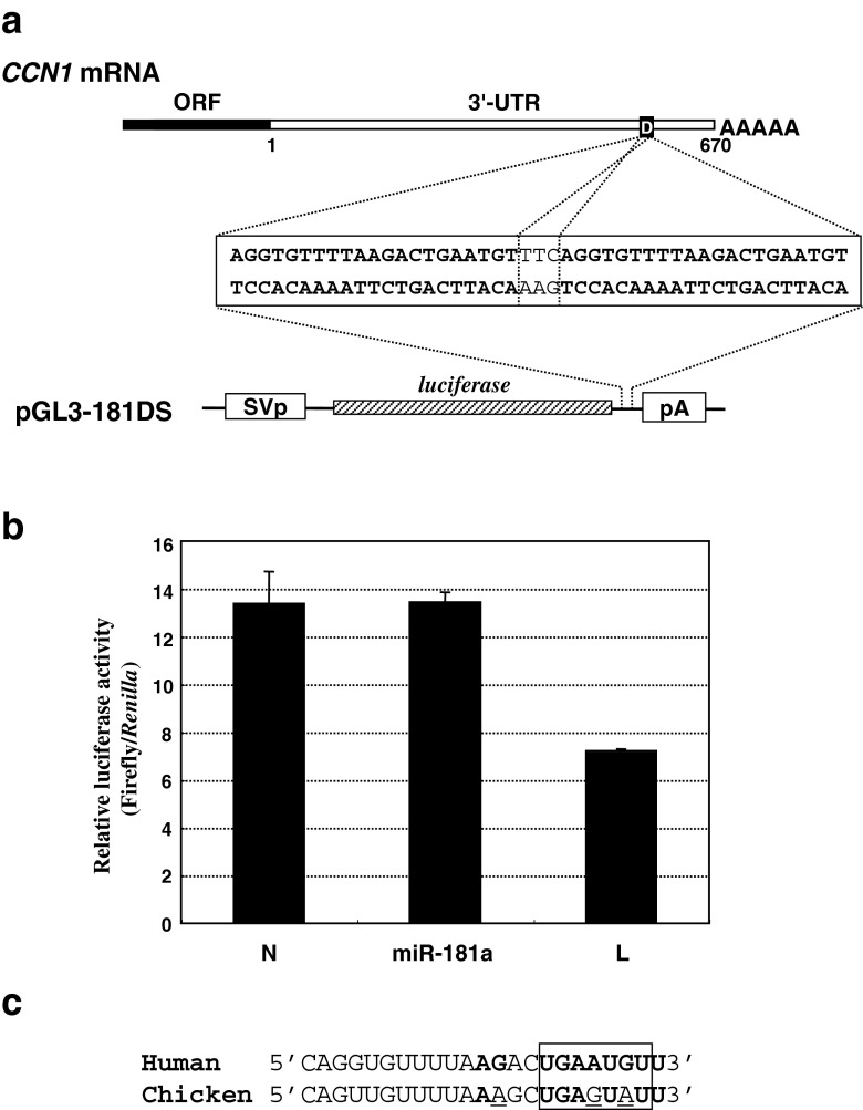
The 3'-untranslated region (UTR) is known to be a critical regulator of post-transcriptional events that determine the gene expression at the RNA level. The gene CCN1 is one of the classical members of the matricellular CCN family and is involved in a number of biological processes during mammalian development. In the present study, the 600-bp 3'-UTR of CCN1 was functionally characterized. Reporter gene analysis revealed that the entire 3'-UTR profoundly repressed gene expression in cis in different types of the cells, to which both the proximal and distal-halves of the 3'-UTR segments contributed almost equally. Deletion analysis of the 3'-UTR indicated a distinct functional element in the proximal half, whereas a putative target for microRNA-181s was predicted in silico in the distal half. Of note, the repressive RNA element in the proximal half was shown to be capable of forming a stable secondary structure. However, unexpectedly, a reporter construct with a tandem repeat of the predicted miR-181 targets failed to respond to miR-181a. In addition, the other major structured element predicted in the distal half was similarly characterized. To our surprise, the second element rather enhanced the reporter gene expression in cis. These results indicate the involvement of multiple regulatory elements in the CCN1 3'-UTR and suggest the complexity of the miRNA action as well as the 3'-UTR-mediated gene regulation.


Figures

Fig. 1
Fig. 1
a Structures of the gene, mRNA, and reporter constructs of human CCN1. The genomic CCN1 structure is illustrated at the top with schematic representation of the corresponding mRNA with 5′-cap and polyadenyl tail. Solid and open boxes denote open reading frames and untranslated regions, respectively, whereas intronic regions are shown in bold lines. In the lower half of the panel, construction of the original reporter plasmids, pGL3-CyrUTRS and pGL3-CyrUTRA, is displayed. SV40p, pA, and 3′-UTR indicate SV40 promoter, SV40 polyadenylation signal, and the 3′-UTR of CCN1, respectively. b Effect of the cis-linked 3′-UTR at the 3′-end on luciferase gene expression. The parental pGL3-L(+) (C), pGL3-CyrUTRS (UTRS), or pGL3-CyrUTRA (UTRA) was used to transfect the cells along with an internal control vector, pRL-TK. Forty-eight hours later, the cells were harvested for the luciferase assay, and firefly luciferase gene activities were standardized against those of the internal control. The name of the cell line used is shown on each graph. Mean values are shown with error bars of standard deviations
Fig. 2
Fig. 2
a Structure of the plasmids used to evaluate the positional effect on the 3′-UTR function. In pGL3-CyrUTR-XN, the CCN1 3′-UTR was relocated immediately upstream of the SV40 promoter. b Structures of the reporter constructs for the independent functional evaluation of the proximal and distal halves of the 3′-UTR. The junction of the cDNA fragments subcloned is indicated by the nucleotide number counted from the upstream end (1) of the 3′-UTR. The small solid box labeled “D” represents the putative miRNA target predicted in silico, which is detailed in panel d. c Relative luciferase activities from the plasmids illustrated in panels a and b in HeLa cells. The left panel shows the results obtained with the plasmids described in panel a, where ∆P indicates a plasmid without a promoter, pGL3∆P. Similarly, the right panel shows those from the plasmids in panel b. Mean values are shown with error bars of standard deviations. d Putative miRNA target in the CCN1 3′-UTR predicted by Targetscan. The nucleotide sequence of the corresponding region in the 3′-UTR is displayed, together with that of miR-181a forming an RNA duplex
Fig. 3
Fig. 3
Post-transcriptional regulatory element in the proximal half of the CCN1 3′-UTR. a Deletion mutants of the 3′-UTR in the reporter gene constructs were made to locate a functional element in the proximal half. Approximate locations and size of the 3′-UTR subfragments are indicated by nucleotide numbers counted from the upstream end of the 3′-UTR. Abbreviations used are as described in Fig. 1 legend. b Relative luciferase activities from the plasmids illustrated in panel a. Evaluation was performed in HeLa cells with a control experiment (C) using pGL3-L(+). Mean values are shown with error bars of standard deviations. c Predicted secondary structure of the minimal functional segment involved in pGL3-CyrUTR-BEMI. The prediction was performed by use of GENETYX software
Fig. 4
Fig. 4
Functional evaluation of the predicted miR-181a target sequence in the CCN1 3′-UTR. a Structure of the plasmid used to evaluate the function of the miR-181a target predicted by Targetscan. A double strand DNA containing 2 copies of the miR-181a target sequences predicted in human CCN1 was synthesized and inserted into the parental reporter plasmid to yield pGL3-181DS. The nucleotide sequence of the corresponding region is illustrated in the middle of the panel. b Relative luciferase activities from the plasmid illustrated in a in HCS-2/8 cells in the presence or absence of exogenous miR-181a. The cells were transfected with miR-181a or a control siRNA (N, negative control siRNA; L, control luciferase siRNA). Eight hours later, the cells were further transfected with the reporter plasmid along with an internal control, phR-TK (int-), and were harvested for the luciferase assay after 48 h of incubation. c Absence of the seed element for miR-181 in chicken CCN1. Nucleotide sequences of the predicted miR-181a target in human CCN1 and the correspondent in chicken CCN1 are aligned. Nucleotides complementary to miR-181a are shown in bold cases, whereas those mismatched in chicken CCN1 are underlined. The seed element is indicated by a box
Fig. 5
Fig. 5
a Distribution of putative structured elements in the 3′-UTR of human CCN1 mRNA. Two major stem-loops and 4 minor ones are represented by red and blue lines, respectively. Functional evaluation firmly indicated the repressive regulatory function of the major stem-loop 1 in the proximal half, which involved the minimal functional segment (BEMI)
Fig. 6
Fig. 6
Functional characterization of the major stem-loop 2 predicted in the distal half of the 3′-UTR. a Structure of the reporter construct. A cDNA corresponding to the major stem-loop 2 illustrated in Fig. 5 was isolated by PCR and was subcloned into the same parental vector at the downstream of the luciferase gene. b Relative luciferase activity in the cell lysate of HeLa cells transfected with the plasmid shown in panel a as represented by the percentage against the control. The parental pGL-3L(+) was utilized for the control experiment
Fig. 7
Fig. 7
Structural characteristics of cis-repressive elements in the 3′-UTR of the CCN family member genes. a Nucleotide sequence alignment of the 3′-UTR segment around the human BEMI element with its corresponding region in chicken CCN1 3′-UTR. Nucleotides matched between the two are shown in bold letters, whereas the BEMI element is underlined. b Predicted secondary structure of the 3′-UTR in chicken CCN1 mRNA. The major and minor stem-loops are represented in red and blue, respectively. The substructure in the major loop (purple) indicates a putative repressive element. c Comparison of the secondary structures of functional cis-repressive elements identified in the 3′-UTRs of CCN family member genes. In addition to the BEMI element found in this study, human and murine cis-acting elements of structure-anchored repression (CAESARs) are illustrated (Kubota et al. ; Kondo et al. 2000)

Similar articles

Cited by

References

    1. Andreassi S, Riccio A. To localize or not to localize: mRNA fate is in 3'UTR ends. Trends Cell Biol. 2009;19:465–474. doi: 10.1016/j.tcb.2009.06.001. - DOI - PubMed
    1. Bai T, Chen CC, Lau LF. Matricellular protein CCN1 activates a proinflammatory genetic program in murine macrophages. J Immunol. 2010;184:3223–3232. doi: 10.4049/jimmunol.0902792. - DOI - PMC - PubMed
    1. Didiano D, Hobert O. Molecular architecture of a miRNA-regulated 3' UTR. RNA. 2008;14:1297–1316. doi: 10.1261/rna.1082708. - DOI - PMC - PubMed
    1. Holbourn KP, Acharya KR, Perbal B. The CCN family of proteins: structure-function relationships. Trends Biochem Sci. 2008;33:461–473. doi: 10.1016/j.tibs.2008.07.006. - DOI - PMC - PubMed
    1. Holloway SE, Beck AW, Girard L, Jaber MR, Barnett CC, Jr, Brekken RA, Fleming JB. Increased expression of Cyr61 (CCN1) identified in peritoneal metastases from human pancreatic cancer. J Am Coll Surg. 2005;200:371–377. doi: 10.1016/j.jamcollsurg.2004.10.005. - DOI - PubMed