. 2013 Aug;94(Pt 8):1791-1806.
doi: 10.1099/vir.0.053686-0. Epub 2013 May 15.

Proposals for the classification of human rhinovirus species A, B and C into genotypically assigned types

Affiliations

Proposals for the classification of human rhinovirus species A, B and C into genotypically assigned types

Chloe L McIntyre et al. J Gen Virol. 2013 Aug.

Abstract

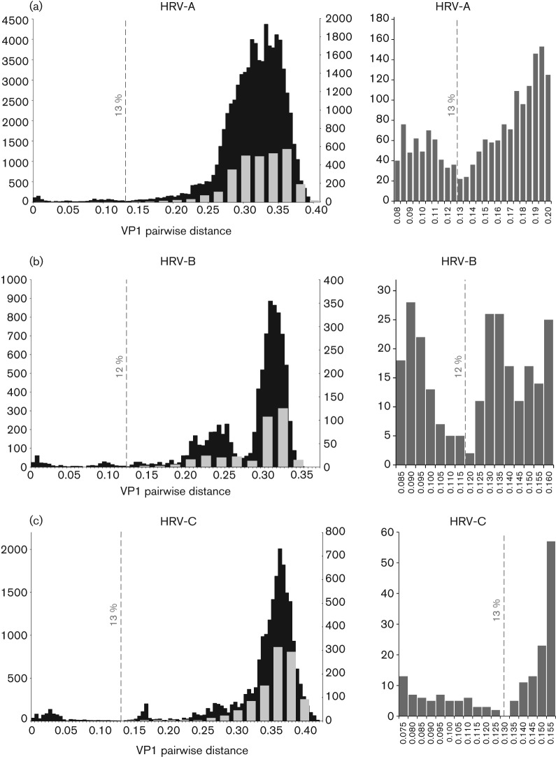
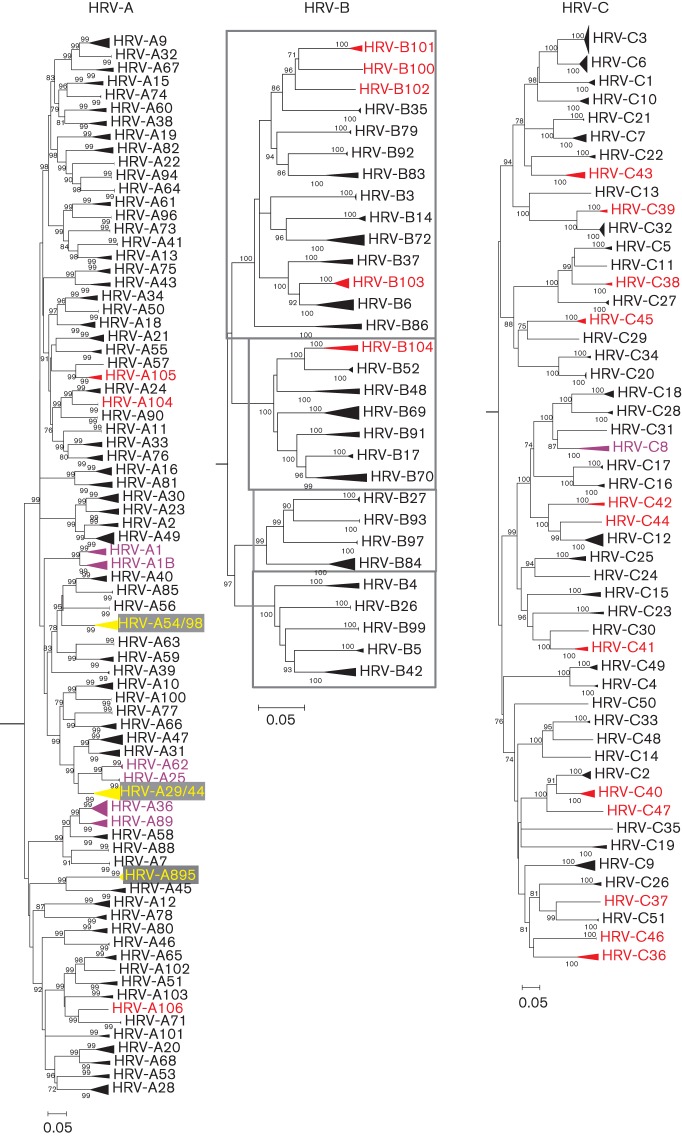
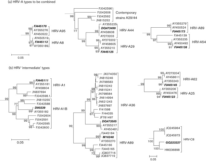
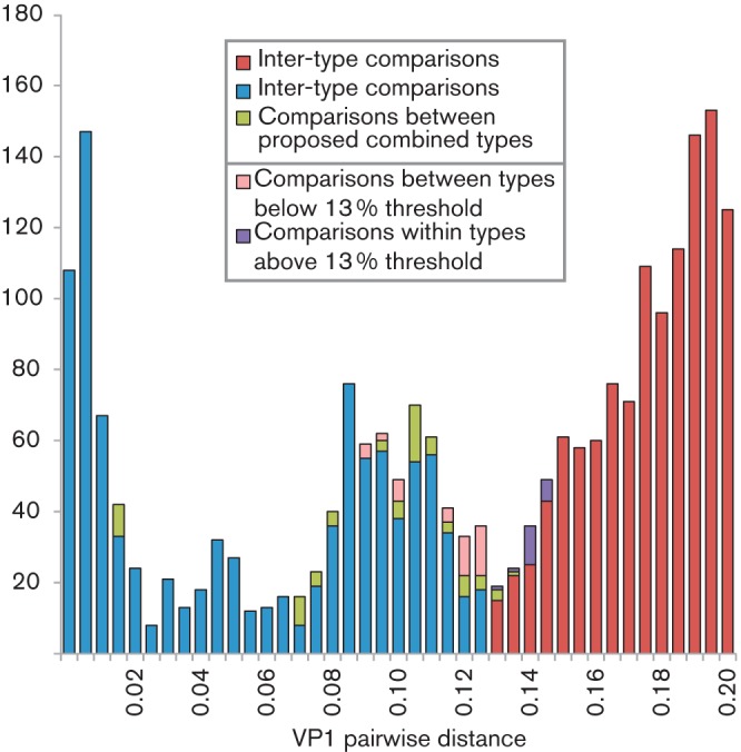
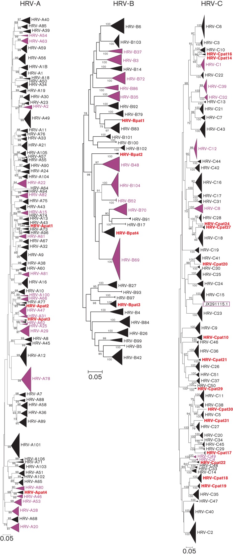
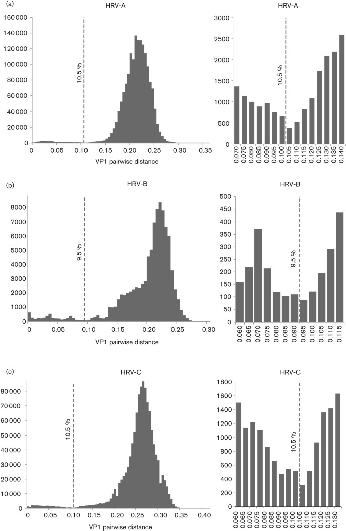
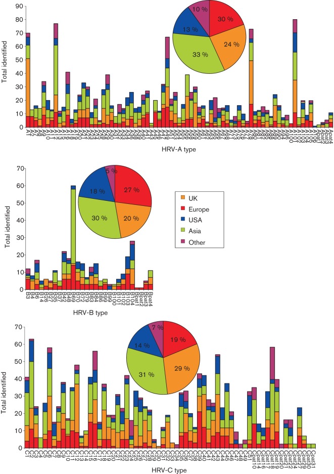
Human rhinoviruses (HRVs) frequently cause mild upper respiratory tract infections and more severe disease manifestations such as bronchiolitis and asthma exacerbations. HRV is classified into three species within the genus Enterovirus of the family Picornaviridae. HRV species A and B contain 75 and 25 serotypes identified by cross-neutralization assays, although the use of such assays for routine HRV typing is hampered by the large number of serotypes, replacement of virus isolation by molecular methods in HRV diagnosis and the poor or absent replication of HRV species C in cell culture. To address these problems, we propose an alternative, genotypic classification of HRV-based genetic relatedness analogous to that used for enteroviruses. Nucleotide distances between 384 complete VP1 sequences of currently assigned HRV (sero)types identified divergence thresholds of 13, 12 and 13 % for species A, B and C, respectively, that divided inter- and intra-type comparisons. These were paralleled by 10, 9.5 and 10 % thresholds in the larger dataset of >3800 VP4 region sequences. Assignments based on VP1 sequences led to minor revisions of existing type designations (such as the reclassification of serotype pairs, e.g. A8/A95 and A29/A44, as single serotypes) and the designation of new HRV types A101-106, B101-103 and C34-C51. A protocol for assignment and numbering of new HRV types using VP1 sequences and the restriction of VP4 sequence comparisons to type identification and provisional type assignments is proposed. Genotypic assignment and identification of HRV types will be of considerable value in the future investigation of type-associated differences in disease outcomes, transmission and epidemiology.


Figures

Fig. 1.
Fig. 1.
Distributions of pairwise nucleotide p-distances for the VP1 region of HRV-A (a), -B (b) and -C (c). Left graphs show number of sequences of the pairwise distances between all available VP1 sequences in black (left y-axis) and a comparison of prototype strains only in grey (right y-axis). The proposed thresholds for type assignment are shown as dotted lines. Right graphs show the sequences distances around the proposed assignment thresholds for each species shown using an expanded x-axis scale.
Fig. 2.
Fig. 2.
Neighbour-joining phylogenetic trees showing the VP1 region of HRV-A, -B and -C. The tree was constructed from by a neighbour-joining method from 100 bootstrap resampled sequence alignments of maximum composite likelihood distances. Multiple sequences of the same type are shown as triangles with heights proportional their numbers and depths corresponding to their earliest diverging branch. Within HRV-B, phylogenetic clusters with pairwise p-distances ranging from 0.1900 to 0.2600 are indicated by grey-outlined boxes. Instances where two types have been combined to form a single type are indicated in yellow. New HRV types defined on the basis of sequence divergence in VP1 are shown in red and HRV types with VP1 intra- or inter-type divergence outside the proposed thresholds are shown in purple. Bars, 0.05 nt substitutions per site.
Fig. 3.
Fig. 3.
Neighbour-joining phylogenetic trees of HRV-A type pairs that do not conform to the proposed VP1 divergence thresholds. Sequences of previously recognized prototype isolates of each HRV type are highlighted in bold and italic. (a) Three HRV-A type pairs that have been combined. (b) Four HRV type pairs that displayed intermediate divergence values are shown. Bars, 0.05 nt substitutions per site.
Fig. 4.
Fig. 4.
Distribution of inter- and intra-type HRV-A pairwise p-distances around the proposed 13 % VP1 divergence threshold. VP1 p-distances between or within types that are consistent with the proposed 13 % type-assignment threshold for VP1 are shown in blue (intra-type) and red (inter-type). Pairwise distances between types that we propose should be combined are shown in green. Comparisons that violate the VP1 assignment threshold are shown in pink (different HRV types but with distances falling below the threshold) and purple (intra-type comparisons falling above the divergence threshold). The y-axis shows the number of sequences.
Fig. 5.
Fig. 5.
Phylogenetic analysis of all available VP4/VP2 sequences of HRV-A, -B and -C. The tree was constructed as described for Fig. 2. Multiple sequences of the same type have been depicted as in Fig. 2. Groups that have been temporarily designated provisionally assigned types (PATs) are shown in red. HRV types that do not conform to the proposed VP4/VP2 thresholds are shown in purple. The single recombinant sequence detected, JX291115, is indicated by a purple box. Bars, 0.05 nt substitutions per site.
Fig. 6.
Fig. 6.
Distributions of pairwise nucleotide p-distances for the VP4/VP2 region of HRV-A, -B and -C. The figure is labelled as in Fig. 1.
Fig. 7.
Fig. 7.
Detection frequencies of different HRV species and types reported from different geographical locations. Total numbers of sequences from each geographical area are shown in the inset pie chart. This analysis excluded HRV types for which geographical information was limited or unavailable (HRV-A50, -A57, -A64, -B5, -B17, -B91 and -C50).

References

    1. Arden K. E., McErlean P., Nissen M. D., Sloots T. P., Mackay I. M. (2006). Frequent detection of human rhinoviruses, paramyxoviruses, coronaviruses, and bocavirus during acute respiratory tract infections. J Med Virol 78, 1232–1240 10.1002/jmv.20689 - DOI - PMC - PubMed
    1. Blomqvist S., Savolainen-Kopra C., Paananen A., Hovi T., Roivainen M. (2009). Molecular characterization of human rhinovirus field strains isolated during surveillance of enteroviruses. J Gen Virol 90, 1371–1381 10.1099/vir.0.008508-0 - DOI - PubMed
    1. Bochkov Y. A., Palmenberg A. C., Lee W.-M., Rathe J. A., Amineva S. P., Sun X., Pasic T. R., Jarjour N. N., Liggett S. B., Gern J. E. (2011). Molecular modeling, organ culture and reverse genetics for a newly identified human rhinovirus C. Nat Med 17, 627–632 10.1038/nm.2358 - DOI - PMC - PubMed
    1. Brown B. A., Maher K., Flemister M. R., Naraghi-Arani P., Uddin M., Oberste M. S., Pallansch M. A. (2009). Resolving ambiguities in genetic typing of human enterovirus species C clinical isolates and identification of enterovirus 96, 99 and 102. J Gen Virol 90, 1713–1723 10.1099/vir.0.008540-0 - DOI - PubMed
    1. Cooney M. K., Fox J. P., Kenny G. E. (1982). Antigenic groupings of 90 rhinovirus serotypes. Infect Immun 37, 642–647 - PMC - PubMed

Associated data