. 2013 Aug;94(Pt 8):1734-1738.
doi: 10.1099/vir.0.053025-0. Epub 2013 May 15.

The respiratory syncytial virus nucleoprotein-RNA complex forms a left-handed helical nucleocapsid

Affiliations

The respiratory syncytial virus nucleoprotein-RNA complex forms a left-handed helical nucleocapsid

Saskia E Bakker et al. J Gen Virol. 2013 Aug.

Abstract

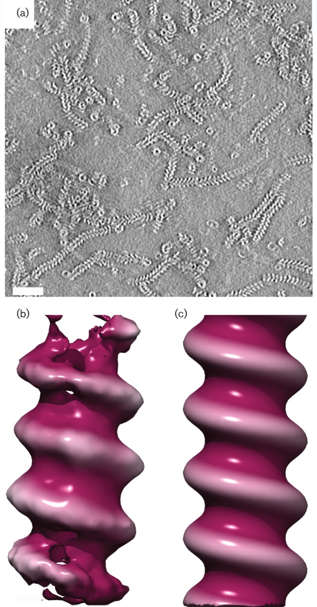
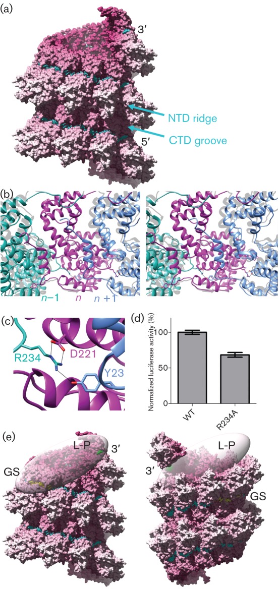
Respiratory syncytial virus (RSV) is an important human pathogen. Its nucleocapsid (NC), which comprises the negative sense RNA viral genome coated by the viral nucleoprotein N, is a critical assembly that serves as template for both mRNA synthesis and genome replication. We have previously described the X-ray structure of an NC-like structure: a decameric ring formed of N-RNA that mimics one turn of the helical NC. In the absence of experimental data we had hypothesized that the NC helix would be right-handed, as the N-N contacts in the ring appeared to more easily adapt to that conformation. We now unambiguously show that the RSV NC is a left-handed helix. We further show that the contacts in the ring can be distorted to maintain key N-N-protein interactions in a left-handed helix, and discuss the implications of the resulting atomic model of the helical NC for viral replication and transcription.


Figures

Fig. 1.
Fig. 1.
Tomography of RSV nucleocapsid. (a) A central section through a denoised tomogram of RSV NC-like RNPs reveals the presence of both rings and short helices. (b) Isosurfaced views of subtomogram averages for RSV nucleocapsid-like RNP reveal that these assemblies are left-handed helices. As the reconstructions did not have sufficient resolution to determine the twist (number of subunits per helical turn), symmetry was imposed. (c) Symmetrized RNP from RSV N-RNA.
Fig. 2.
Fig. 2.
Modelling the RSV nucleocapsid. (a) The left-handed helix obtained by maximizing the preservation of key N–N interactions observed in the ring. A surface rendering of the helix is shown in which the RNA is represented by cyan sticks. (b) Stereo view of the N–N interactions in the modelled helix. The N-terminal arm of subunit n+1 (blue) binds in the core of subunit n (magenta). The ribbon structure for three subunits from the ring was aligned to subunit n and is shown in grey. (c) The inter-subunit interactions are mediated by hydrogen bonds between residue Asp221 of subunit n, Tyr23 of subunit n+1, and Arg234 of subunit n−1 (coloured as in panel b). (d) Evaluation of the effect of R234A mutation on viral RNA synthesis using an RSV based minigenome assay revealed that the mutant N-protein reduced polymerase activity to 68±8 % of the WT level. (e) Implications of the left-hand versus right-handed helical NC for transcription initiation. The left-handed and right-handed NC helix models are shown in surface representation, with the RNA in cyan sticks, except for the nucleotides corresponding to the leader sequence at the extreme 3′ end of the genome and the first gene-start element, shown in green and yellow, respectively. The two domains of the N-protein, NTD and CTD, are such that adjacent CTDs form a continuous groove along the helix, and the NTDs form a helical ridge, labelled in the figure. In the left-handed NC, the bulky NTD projects away from the polymerase complex, allowing simultaneous access to both sequence elements, whereas in the right-handed NC helix, it would project toward the polymerase, interfering with access to the gene-start element.

Similar articles

Cited by

References

    1. Bharat T. A., Riches J. D., Kolesnikova L., Welsch S., Krähling V., Davey N., Parsy M. L., Becker S., Briggs J. A. (2011). Cryo-electron tomography of Marburg virus particles and their morphogenesis within infected cells. PLoS Biol 9, e1001196 10.1371/journal.pbio.1001196 - DOI - PMC - PubMed
    1. Bhella D., Ralph A., Murphy L. B., Yeo R. P. (2002). Significant differences in nucleocapsid morphology within the Paramyxoviridae. J Gen Virol 83, 1831–1839 - PubMed
    1. Bhella D., Ralph A., Yeo R. P. (2004). Conformational flexibility in recombinant measles virus nucleocapsids visualised by cryo-negative stain electron microscopy and real-space helical reconstruction. J Mol Biol 340, 319–331 10.1016/j.jmb.2004.05.015 - DOI - PubMed
    1. Castaño-Díez D., Kudryashev M., Arheit M., Stahlberg H. (2012). Dynamo: a flexible, user-friendly development tool for subtomogram averaging of cryo-EM data in high-performance computing environments. J Struct Biol 178, 139–151 10.1016/j.jsb.2011.12.017 - DOI - PubMed
    1. Egelman E. H., Wu S. S., Amrein M., Portner A., Murti G. (1989). The Sendai virus nucleocapsid exists in at least four different helical states. J Virol 63, 2233–2243 - PMC - PubMed

Publication types

Associated data