. 2013 Jun;6(3):279-89.
doi: 10.1161/CIRCGENETICS.113.000138.

Exome sequencing and systems biology converge to identify novel mutations in the L-type calcium channel, CACNA1C, linked to autosomal dominant long QT syndrome

Affiliations

Exome sequencing and systems biology converge to identify novel mutations in the L-type calcium channel, CACNA1C, linked to autosomal dominant long QT syndrome

Nicole J Boczek et al. Circ Cardiovasc Genet. 2013 Jun.

Abstract

Background: Long QT syndrome (LQTS) is the most common cardiac channelopathy with 15 elucidated LQTS-susceptibility genes. Approximately 20% of LQTS cases remain genetically elusive.

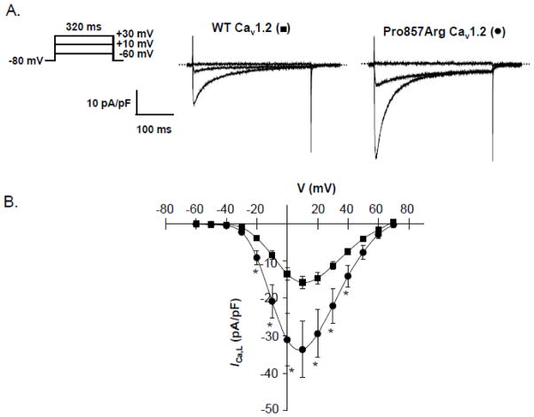
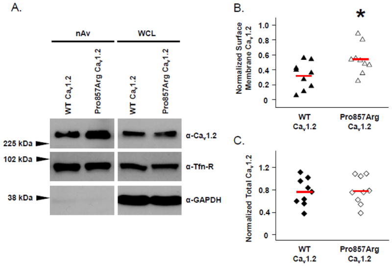
Methods and results: We combined whole-exome sequencing and bioinformatic/systems biology to identify the pathogenic substrate responsible for nonsyndromic, genotype-negative, autosomal dominant LQTS in a multigenerational pedigree, and we established the spectrum and prevalence of variants in the elucidated gene among a cohort of 102 unrelated patients with "genotype-negative/phenotype-positive" LQTS. Whole-exome sequencing was used on 3 members within a genotype-negative/phenotype-positive family. Genomic triangulation combined with bioinformatic tools and ranking algorithms led to the identification of a CACNA1C mutation. This mutation, Pro857Arg-CACNA1C, cosegregated with the disease within the pedigree, was ranked by 3 disease-network algorithms as the most probable LQTS-susceptibility gene and involves a conserved residue localizing to the proline, gltamic acid, serine, and threonine (PEST) domain in the II-III linker. Functional studies reveal that Pro857Arg-CACNA1C leads to a gain of function with increased ICa,L and increased surface membrane expression of the channel compared to wild type. Subsequent mutational analysis identified 3 additional variants within CACNA1C in our cohort of 102 unrelated cases of genotype-negative/phenotype-positive LQTS. Two of these variants also involve conserved residues within Cav1.2's PEST domain.

Conclusions: This study provides evidence that coupling whole-exome sequencing and bioinformatic/systems biology is an effective strategy for the identification of potential disease-causing genes/mutations. The identification of a functional CACNA1C mutation cosegregating with disease in a single pedigree suggests that CACNA1C perturbations may underlie autosomal dominant LQTS in the absence of Timothy syndrome.


Conflict of interest statement

Conflict of Interest Disclosures: M.J.A is a consultant for Transgenomic. Intellectual Property derived from M.J.A’s research program resulted in license agreements in 2004 between Mayo Clinic Health Solutions (formerly Mayo Medical Ventures) and PGxHealth (formerly Genaissance Pharmaceuticals and now Transgenomic). However, Transgenomic did not contribute directly to this study in any manner. NJB, JMB, DJT, JRG, SM, JME, TJK and MJA have no conflicts of interest.

Figures

Figure 1
Figure 1
Whole Exome Sequencing and Familial Genomic Triangulation for the Elucidation of a Novel Genetic Substrate for LQTS. (A) Black circles/squares are affected, grey are borderline, and white are unaffected with LQTS. Arrow identifies the proband. Asterisks represent those who were whole exome sequenced. Numbers in each circle/square represent QTc values (milliseconds) when available. Plus signs represent Pro857Arg-CACNA1C mutation positive family members. Minus signs represent mutation negative family members. NA denotes where DNA samples were not available. (B) Lead II of the electrocardiogram for the index case (III.2). The full electrocardiogram is available in Supplemental Figure 1A. (C) Schematic representation of our sequencing strategy. (D) DNA sequencing chromatogram showing the Pro857Arg mutation.
Figure 2
Figure 2
Functional Analysis of Pro857Arg-Cav1.2 Mutation Using Whole Cell Patch Clamp Technique. (A) Representative ICa,L traces recorded from HEK293 cells expressing WT- or Pro857Arg-Cav1.2 along with auxiliary β2cN4 and α2δ1 subunits. (B) Current-voltage (I–V) relations constructed from WT-Cav1.2 (■, n = 10) or Pro857Arg-Cav1.2 expressing cells (●, n = 10). Data were compared using Student’s unpaired t-test and * denotes p < 0.05 when WT-Cav1.2 was compared with Pro857Arg-Cav1.2.
Figure 3
Figure 3
Increased Surface Membrane Density of Cav1.2 Pro857Arg Channels. HEK293 cells expressing either YFP-tagged Cav1.2 WT or Cav1.2-Pro857Arg along with auxiliary β2cN4 and α2δ1 subunits were cell surface biotinylated at 4°C. (A) NeutrAvidin (nAv)-captured surface membrane proteins (left panels) and whole cell lysates (WCL, right panels) were analyzed by SDS-PAGE and western blotting. Densitometric analysis of surface membrane Cav1.2 signal (B) or total cellular Cav1.2 (C) was performed to summarize the results of nine independent experiments. In both cases, normalization of Cav1.2 signal was achieved using the corresponding signal of the endogenous surface membrane protein transferrin receptor (Tfn-R). Solid red lines through distributions indicate population means. Data were compared using Student’s unpaired t-test and * denotes p ≤ 0.05 when WT-Cav1.2 was compared with Pro857Arg-Cav1.2.
Figure 4
Figure 4
Topology Diagram of the CACNA1C Channel Alpha-Subunit. Each variant is represented as a black circle followed by the amino acid position and change. Asterisk represents the mutation identified by whole exome sequencing in the pedigree (Figure 1). Patient history is displayed for each variant. The PEST domain sequence is highlighted showing the position of the three variants residing within this domain as well as the species conservation. STIM1 binding region, 1806–1905 is shown in the C-terminal tail of CACNA1C. STIM1 interacts with the LTCC to normally suppress channel function , .

Comment in

References

    1. Liberthson RR. Sudden death from cardiac causes in children and young adults. N Engl J Med. 1996;334:1039–1044. - PubMed
    1. Chugh SS, Reinier K, Teodorescu C, Evanado A, Kehr E, Al Samara M, et al. Epidemiology of sudden cardiac death: Clinical and research implications. Prog Cardiovasc Dis. 2008;51:213–228. - PMC - PubMed
    1. Tester DJ, Ackerman MJ. Postmortem long qt syndrome genetic testing for sudden unexplained death in the young. J Am Coll Cardiol. 2007;49:240–246. - PubMed
    1. Schwartz PJ, Stramba-Badiale M, Crotti L, Pedrazzini M, Besana A, Bosi G, et al. Prevalence of the congenital long-qt syndrome. Circulation. 2009;120:1761–1767. - PMC - PubMed
    1. Moss AJ, Kass RS. Long qt syndrome: From channels to cardiac arrhythmias. J Clin Invest. 2005;115:2018–2024. - PMC - PubMed

Publication types

Substances