. 2013 Jun 21;288(25):18249-59.
doi: 10.1074/jbc.M112.446385. Epub 2013 May 15.

Nilotinib induces autophagy in hepatocellular carcinoma through AMPK activation

Affiliations

Nilotinib induces autophagy in hepatocellular carcinoma through AMPK activation

Hui-Chuan Yu et al. J Biol Chem. .

Abstract

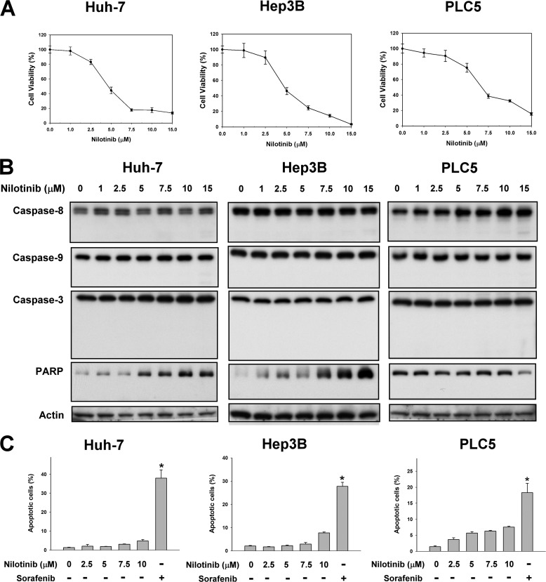
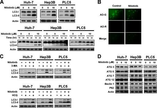
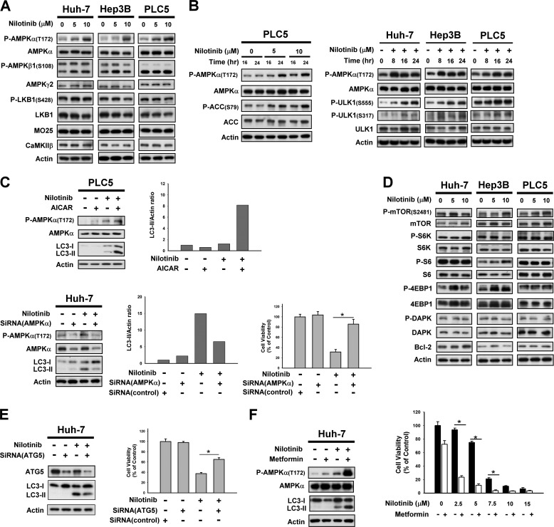
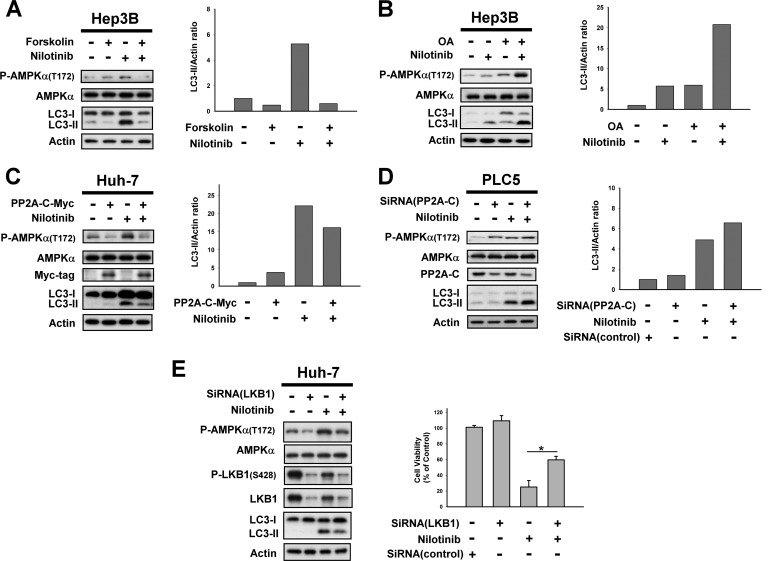
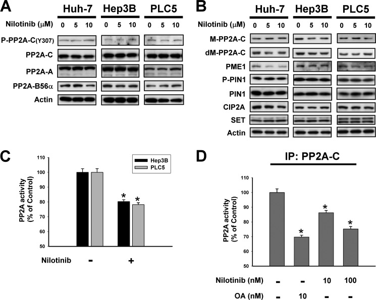
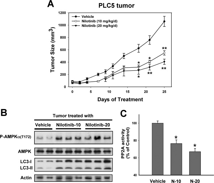
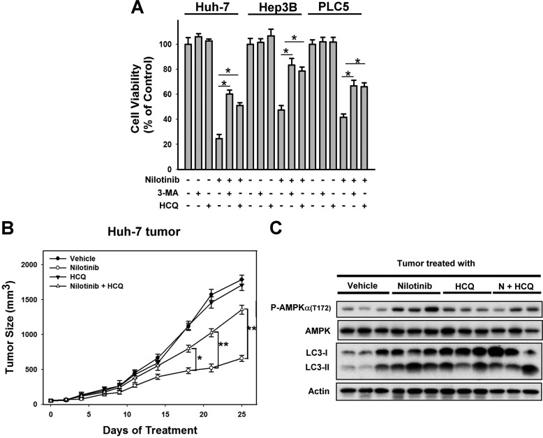
Hepatocellular carcinoma (HCC) is the most common liver cancer and the third-leading cause of cancer death worldwide. Nilotinib is an orally available receptor tyrosine kinase inhibitor approved for chronic myelogenous leukemia. This study investigated the effect of nilotinib on HCC. Nilotinib did not induce cellular apoptosis. Instead, staining with acridine orange and microtubule-associated protein 1 light chain 3 revealed that nilotinib induced autophagy in a dose- and time-dependent manner in HCC cell lines, including PLC5, Huh-7, and Hep3B. Moreover, nilotinib up-regulated the phosphryaltion of AMP-activated kinase (AMPK) and protein phosphatase PP2A inactivation were detected after nilotinib treatment. Up-regulating PP2A activity suppressed nilotinib-induced AMPK phosphorylation and autophagy, suggesting that PP2A mediates the effect of nilotinib on AMPK phosphorylation and autophagy. Our data indicate that nilotinib-induced AMPK activation is mediated by PP2A, and AMPK activation and subsequent autophagy might be a major mechanism of action of nilotinib. Growth of PLC5 tumor xenografts in BALB/c nude mice was inhibited by daily oral treatment with nilotinib. Western blot analysis showed both increased phospho-AMPK expression and decreased PP2A activity in vivo. Together, our results reveal that nilotinib induces autophagy, but not apoptosis in HCC, and that the autophagy-inducing activity is associated with PP2A-regulated AMPK phosphorylation.

Keywords: AMP-activated Kinase (AMPK); Apoptosis; Autophagy; HCC; Nilotinib; Serine Threonine Protein Phosphatase; Tyrosine Protein Kinase (Tyrosine Kinase).


Figures

FIGURE 1.
FIGURE 1.
Nilotinib reduced cell viability in HCC but did not induce apoptosis. A, dose-dependent effects of nilotinib on cell viability. HCC cells were treated with nilotinib at the indicated concentrations for 72 h. Cell viability was measured by MTT assay. Points, mean; bars, S.D. (n = 8). B and C, effects of nilotinib on apoptosis. B, cells were exposed to nilotinib at the indicated concentrations for 48 h. Cell lysates were assayed by Western blot to detect the activation of caspases and PARP cleavage. C, apoptotic cells (sub-G1) were analyzed by flow cytometry after cells were exposed to nilotinib at the indicated concentrations or sorafenib at 20 μm for 24 h.
FIGURE 2.
FIGURE 2.
Nilotinib induces autophagy in HCC. A, top, dose-dependent effects of nilotinib-induced autophagy. HCC cells were treated with nilotinib at the indicated concentrations for 24 h. Bottom, time-dependent effects of nilotinib-induced autophagy. Cells were treated with nilotinib at the indicated concentrations for 16 or 24 h. Cell lysates were prepared for immunoblotting of microtubule-associated protein 1 light chain 3 (LC3). B, LC3-II immunofluorescence and acridine orange stain. Hep3B cells were treated with nilotinib at 10 μm for 24 h. C, top, co-treatment with autophagy inhibitor 3-MA reduced nilotinib-induced autophagy. Cells were treated with nilotinib (10 μm) and/or 3-MA (1 mm) for 24 h. Bottom, co-treatment with HCQ reduced nilotinib-induced autophagy. Cells were treated with nilotinib (10 μm) and/or HCQ (10 μm) for 24 h. D, dose-dependent analysis of autophagy-related proteins. Cells were exposed to nilotinib at the indicated concentrations for 24 h.
FIGURE 3.
FIGURE 3.
AMPK mediated nilotinib-induced autophagy. A, target validation of CIP2A-Akt-4EBP1. A, dose-dependent analysis of AMPK-related molecules in HCC cells. Cells were exposed to nilotinib at the indicated concentrations for 24 h. B, time-dependent analysis of P-AMPKα, AMPKα, P-ACC, ACC, P-ULK1, and ULK1 in HCC cells. Cells were exposed to nilotinib at the indicated concentrations for 24 h. C, validation of AMPKα mediation of nilotinib-induced autophagy. Top, co-treatment with AICAR, an activator of AMPK, increased nilotinib-induced autophagy. Cells were treated with nilotinib (10 μm) and/or AICAR (1 mm) for 24 h. Bottom, silencing AMPKα by siRNA reduced nilotinib-induced autophagy and cell death. Huh-7cells were transfected with control or AMPKα siRNA for 48 h then treated with nilotinib (10 μm) for 24 h. Cell viability was measured by MTT assay. D, dose-dependent analysis of other autophagy-related molecules. Cells were exposed to nilotinib at the indicated concentrations for 24 h. E, silencing Atg 5 by siRNA reduced nilotinib-induced autophagy and cell death. Huh-7 cells were transfected with siRNA for 48 h then treated with nilotinib (10 μm) for 24 h. Cell viability was measured by MTT assay. F, adding metformin increased nilotinib-induced autophagy and cell death. Cells were exposed to nilotinib at 10 μm or at the indicated concentrations and/or metformin at 10 mm for 24 h.
FIGURE 4.
FIGURE 4.
PP2A mediates nilotinib-induced activation of AMPK. A, left, co-treatment with forskolin, a PP2A agonist, reverses the effect of nilotinib on P-AMPKα and autophagy. Right, immunoblots were scanned using a UVP BioSpectrum AC image system and quantitated using VisionWork LS software to determine the ratio of the level of LC3-II to actin. B, co-treatment with okadaic acid, a PP2A inhibitor, enhanced the effect of nilotinib on P-AMPKα and autophagy. C, ectopic expression of AMPKα abolishes effects of nilotinib on autophagy in Huh-7 cells. Cells were transfected with AMPKα-myc and were selected for 8 weeks by G-418. Analysis of autophagy was performed by Western blot (WB) after cells were sequentially exposed to DMSO or nilotinib (10 μm) for 24 h. D, knockdown of PP2A-C enhanced nilotinib-induced autophagy. PLC5 cells were transfected with control or AMPKα siRNA for 48 h and then treated with nilotinib (10 μm) for 24 h. E, silencing LKB1 by siRNA reduced nilotinib-induced autophagy. Huh-7 cells were transfected with control or LKB1 siRNA for 48 h then treated with nilotinib (10 μm) for 24 h. Cell viability was measured by MTT assay.
FIGURE 5.
FIGURE 5.
Nilotinib reduced the activity of PP2A. A, dose-dependent effects of nilotinib on P-PP2A-C, PP2A-C, PP2A-A, and PP2A-B56α. B, dose-dependent effects of nilotinib on PP2A-related proteins. C, treatment of nilotinib reduced the activity of PP2A. D, effects of nilotinib on PP2A activity in PP2A-C-containing lysates. Hep3B cells were immunoprecipitated with anti-PP2A-C then incubated with drugs for 24 h. Columns, mean; bars, S.D. (n = 3). *, p < 0.05.
FIGURE 6.
FIGURE 6.
In vivo effect of nilotinib on PLC5 xeonograft nude mice. A, tumor growth curves of PLC5. Points, mean (n = 6); bars, S.E. *, p < 0.05; **, p < 0.01. B, Western blot analysis of P-AMPK, AMPK, and LC-3 in PLC5 tumors. C, analysis of PP2A activity. Columns, mean; bars, S.D. (n = 6). *, p < 0.05 versus vehicle group.
FIGURE 7.
FIGURE 7.
Inhibition of autophagy reduced the anti-tumor effect of nilotinib in vitro and in vivo. A, co-treatment with autophagy inhibitors, 3-methyladenine (3-MA, 1 mm) or hydroxychloroquine (HCQ, 10 μm), reduced the effect of nilotinib on cell death. Cells were treated with nilotinib at10 μm and/or 3-MA/HCQ for 24 h. Cell viability was analyzed by MTT assay. B, combination of nilotinib and HCQ reduced the effect of nilotinib on Huh-7 tumors. Points, mean (n = 6); bars, S.E. *, p < 0.05; **, p < 0.01. (nilotinib group versus nilotinib plus HCQ group). C, Western blot analysis of P-AMPK, AMPK, and LC-3 in Huh-7 tumors.

References

    1. Adnane L., Trail P. A., Taylor I., Wilhelm S. M. (2006) Sorafenib (BAY 43–9006, Nexavar), a dual-action inhibitor that targets RAF/MEK/ERK pathway in tumor cells and tyrosine kinases VEGFR/PDGFR in tumor vasculature. Methods Enzymol. 407, 597–612 - PubMed
    1. Al-Kandari F. A., Owunwanne A., Syed G. M., Ar Marouf R., Elgazzar A. H., Shiekh M., Rizui A. M., Al-Ajmi J. A., Mohammed A. M. (2007) Regional cerebral blood flow in patients with sickle cell disease: study with single photon emission computed tomography. Ann. Nucl. Med. 21, 439–445 - PubMed
    1. Auclair D., Miller D., Yatsula V., Pickett W., Carter C., Chang Y., Zhang X., Wilkie D., Burd A., Shi H., Rocks S., Gedrich R., Abriola L., Vasavada H., Lynch M., Dumas J., Trail P. A., Wilhelm S. M. (2007) Antitumor activity of sorafenib in FLT3-driven leukemic cells. Leukemia 21, 439–445 - PubMed
    1. Wilhelm S., Carter C., Lynch M., Lowinger T., Dumas J., Smith R. A., Schwartz B., Simantov R., Kelley S. (2006) Discovery and development of sorafenib: a multikinase inhibitor for treating cancer. Nat. Rev. Drug Discov. 5, 835–844 - PubMed
    1. Cheng A. L., Kang Y. K., Chen Z., Tsao C. J., Qin S., Kim J. S., Luo R., Feng J., Ye S., Yang T. S., Xu J., Sun Y., Liang H., Liu J., Wang J., Tak W. Y., Pan H., Burock K., Zou J., Voliotis D., Guan Z. (2009) Efficacy and safety of sorafenib in patients in the Asia-Pacific region with advanced hepatocellular carcinoma: a phase III randomized, double-blind, placebo-controlled trial. Lancet Oncol. 10, 25–34 - PubMed

Publication types

MeSH terms

LinkOut - more resources