. 2013 Jul 5;288(27):19343-57.
doi: 10.1074/jbc.M113.479725. Epub 2013 May 15.

Specificity of intersubunit general anesthetic-binding sites in the transmembrane domain of the human α1β3γ2 γ-aminobutyric acid type A (GABAA) receptor

Affiliations

Specificity of intersubunit general anesthetic-binding sites in the transmembrane domain of the human α1β3γ2 γ-aminobutyric acid type A (GABAA) receptor

David C Chiara et al. J Biol Chem. .

Abstract

GABA type A receptors (GABAAR), the brain's major inhibitory neurotransmitter receptors, are the targets for many general anesthetics, including volatile anesthetics, etomidate, propofol, and barbiturates. How such structurally diverse agents can act similarly as positive allosteric modulators of GABAARs remains unclear. Previously, photoreactive etomidate analogs identified two equivalent anesthetic-binding sites in the transmembrane domain at the β(+)-α(-) subunit interfaces, which also contain the GABA-binding sites in the extracellular domain. Here, we used R-[(3)H]5-allyl-1-methyl-5-(m-trifluoromethyl-diazirynylphenyl) barbituric acid (R-mTFD-MPAB), a potent stereospecific barbiturate anesthetic, to photolabel expressed human α1β3γ2 GABAARs. Protein microsequencing revealed that R-[(3)H]mTFD-MPAB did not photolabel the etomidate sites at the β(+)-α(-) subunit interfaces. Instead, it photolabeled sites at the α(+)-β(-) and γ(+)-β(-) subunit interfaces in the transmembrane domain. On the (+)-side, α1M3 was labeled at Ala-291 and Tyr-294 and γ2M3 at Ser-301, and on the (-)-side, β3M1 was labeled at Met-227. These residues, like those in the etomidate site, are located at subunit interfaces near the synaptic side of the transmembrane domain. The selectivity of R-etomidate for the β(+)-α(-) interface relative to the α(+)-β(-)/γ(+)-β(-) interfaces was >100-fold, whereas that of R-mTFD-MPAB for its sites was >50-fold. Each ligand could enhance photoincorporation of the other, demonstrating allosteric interactions between the sites. The structural heterogeneity of barbiturate, etomidate, and propofol derivatives is accommodated by varying selectivities for these two classes of sites. We hypothesize that binding at any of these homologous intersubunit sites is sufficient for anesthetic action and that this explains to some degree the puzzling structural heterogeneity of anesthetics.

Keywords: Anesthetics; Barbiturates; Cys Loop Receptors; Etomidate; GABA Receptors; Nicotinic Acetylcholine Receptors; Photoaffinity Labeling; Propofol.


Figures

FIGURE 1.
FIGURE 1.
Locations in an (α)2(β)2γ GABAAR of binding sites for GABA, benzodiazepines (BZD), and etomidate.
FIGURE 2.
FIGURE 2.
R- [3H]mTFD-MPAB and R- [3H]azietomidate photolabeling of α1β3 and α1β3γ2 GABAARs. GABAARs were photolabeled with 0.8 μm R-[3H]azietomidate or 0.3 μm R-[3H]mTFD-MPAB in the absence and presence of 1 mm pentobarbital (PB), and aliquots (∼5 pmol of [3H]muscimol sites/lane) were fractionated by SDS-PAGE. A, Coomassie Blue (Coo Blue) stain of representative gel lanes, with the mobilities of molecular weight markers and the calculated masses of the stained gel bands indicated. B, 3H incorporation into GABAAR subunits, as determined by fluorography (1 month exposure). C, quantitation of the α1, β3, and γ2 subunit distributions, as determined by Edman sequence analysis of materials extracted from the stained gel bands. α1 was concentrated in the 56-kDa band and β3 in the 59- and 61-kDa bands. The γ2 subunit is distributed diffusely in all three bands.
FIGURE 3.
FIGURE 3.
R-[3H]mTFD-MPAB binds to site(s) in the α1β3γ2 GABAAR that are distinct from, but coupled energetically to, the etomidate and GABA-binding sites. GABAARs in the absence (●, ○, ■) or presence (□) of GABA were photolabeled on an analytical scale with R-[3H]mTFD-MPAB (A, C, and E) or R-[3H]azietomidate (B, D, and F) in the presence of increasing concentrations of nonradioactive R-mTFD-MPAB (●) or S-mTFD-MPAB (○) (−GABA; A and B), R-etomidate (C and D), or pentobarbital (E and F), and 3H incorporation into GABAAR subunits was determined by SDS-PAGE and liquid scintillation counting. The concentration dependences of inhibition (IC50) and potentiation (EC50) were fit as described under “Experimental Procedures,” and the values of IC50/EC50 of the plotted lines are included under the “Results.” The amounts of R-[3H]mTFD-MPAB incorporation in the presence of 1 mm pentobarbital or R-[3H]azietomidate incorporation in the presence of 1 mm R-etomidate are indicated by long dashed lines.
FIGURE 4.
FIGURE 4.
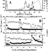
R-[3H]mTFD-MPAB photolabels β3Met-227 in the β3M1 transmembrane helix. GABAARs were photolabeled on a preparative scale in the presence of 1 mm GABA with R-[3H]mTFD-MPAB (0.6 μm) in the absence (●, □) or presence (○) of 1 mm pentobarbital, and GABAAR subunits were isolated by SDS-PAGE. A, rpHPLC fractionation of EndoLys-C digests of β3 subunits (59–61-kDa gel bands). Fractions 28–29 containing the peak of 3H were pooled for sequencing (B). B and C, 3H (●, ○) and picomoles of PTH-derivatives (□) released during Edman sequencing of β3 subunit fragments beginning at β3Arg-216 (B) and β3His-191 (C). B, primary sequence began at β3Arg-216 (35 pmol, both conditions), and the peak of 3H release in cycle 12 indicated photolabeling of β3Met-227 in βM1 at 980 cpm/pmol (−pentobarbital) and 160 cpm/pmol (+pentobarbital). C, aliquots of β3 subunit from the same preparative labeling were digested with EndoGlu-C and sequenced without fractionation. The sequencing filters were treated with OPA prior to cycle 16, and thereafter, the only sequence detected originally began at β3His-191 (4–5 pmol). The peak of 3H release in cycle 37 confirmed R-[3H]mTFD-MPAB photolabeling of β3Met-227 at 920 cpm/pmol in the absence and 170 cpm/pmol in the presence of pentobarbital.
FIGURE 5.
FIGURE 5.
R-[3H]mTFD-MPAB photolabels α1Ala-291, α1Tyr-294, and γ2Ser-301 in the α1 and γ2 M3 transmembrane helices. A, 3H (●) and picomoles of PTH-derivatives (□) released during Edman sequencing of a GABAAR subunit fragment beginning at α1Asp-287 (7 pmol). The peaks of 3H release in cycles 5 and 8 indicated photolabeling of α1Ala-291 and α1Tyr-294. For this sequencing experiment, material isolated by rpHPLC from an EndoGlu-C digest of α1 subunit (B, fractions 25–27) was sequenced for four cycles, establishing that the primary sequence began at α1Ser-251 before α1M2, a fragment predicted to extend to α1Glu-313 near the C terminus of α1M3 (C). After cycle 4, the sample was treated with OPA to block all free N termini, which was confirmed by five more cycles of Edman degradation, and then treated with cyanogen bromide to cleave at methionines before sequencing for 15 additional cycles. D, 3H (●) and picomoles of PTH-derivatives (□) released during Edman sequencing of a GABAAR subunit fragment beginning at γ2Asp-297 (0.6 pmol). The peak of 3H release in cycle 5 indicated labeling of γ2Ser-301. Material isolated by rpHPLC from an EndoGlu-C digest of 59–61-kDa gel bands (E, fractions 28–29)) was sequenced for 10 cycles, establishing the presence of the fragment beginning at γ2Val-212 (F) as a secondary sequence along with the primary sequence beginning at β3His-191. The sample was treated with OPA after cycle 10 to block all free N termini, sequenced an additional 5 cycles to confirm block, then treated with cyanogen bromide, and sequenced for an additional 15 cycles. The efficiencies of photolabeling of the residues are tabulated in Table 5.
FIGURE 6.
FIGURE 6.
R-mTFD-MPAB binds in the GABAAR transmembrane domain to sites at the α+ and γ+ interfaces that are homologous to the etomidate-binding sites at the β+ interfaces. A, side view of an α1β3γ2 GABAAR homology model built using a GLIC crystal structure (Protein Data Bank code 3P50), with α-helices displayed as cylinders, β-sheets as ribbons, and subunits color-coded as follows: α1, light yellow; β3, light blue, and γ2, light green. B and C, views down the ion channel of the GABAAR extracellular (B) and transmembrane (C) domains. A–C, locations are indicated of the pockets containing the binding sites for GABA (green), benzodiazepine (blue), etomidate (brown), and R-mTFD-MPAB (red). D and E, views of R-mTFD-MPAB docked in the pocket at the α+ interface, viewed from the lipid (D) and from the base of the extracellular domain (E). F and G, views from the lipid of R-mTFD-MPAB docked at the γ+ interface (F) and R-azietomidate docked at the β+ interface (G). D–G, docked anesthetic is shown in stick format in its lowest energy orientation, color-coded by element (carbon, gray; oxygen, red; nitrogen, blue; and fluorine, light blue) within the Connolly surface representation of the volumes defined by the ensemble of the 100 lowest energy-minimized docking solutions. Residues photolabeled by R-[3H]mTFD-MPAB are shown in stick format and color-coded as follows: β3Met-227, red; α1Ala-291, magenta; α1Tyr-294, purple; γ2Ser-301, orange; β3Met-286, cyan, and β3Phe-289, yellow-green. Residues photolabeled by R-[3H]azietomidate/[3H]TDBzl-etomidate (G only) are color-coded as follows: β3Met-286, cyan; β3Val-290, dark green; and α1Met-236, lime green. Also color-coded in G are β3Asn-265 (brown, M2–15′), the in vivo etomidate/propofol/pentobarbital sensitivity determinant (5, 6) and β3Phe-301 (blue), the residue photolabeled by an anesthetic steroid in a homopentameric β3 GABAAR (42). The color-coded residues of D–G are also highlighted in the aligned GABAAR subunit sequences spanning the M1–M3 helices (bottom of figure).
FIGURE 7.
FIGURE 7.
Modulation of R-[3H]mTFD-MPAB and R-[3H]azietomidate GABAAR photolabeling by propofol, propofol analogs, alphaxalone, and octanol. α1β3γ2 GABAARs were equilibrated with R-[3H]mTFD-MPAB (A, C, E, and G) or R-[3H]azietomidate (B, D, F, and H) in the absence (●) or presence (▿) of GABA and varying concentrations of propofol (A and B), 2,6-di-sec-butylphenol (○) or 2,6-di-tert-butylphenol (♢) (C and D (+GABA)), alphaxalone (E and F), or octanol (G and H). The concentration dependences of potentiation (EC50) and inhibition (IC50) were fit as described under “Experimental Procedures,” and the values of IC50/EC50 of the plotted lines are included under the “Results.” For each experiment, the amounts of R-[3H]mTFD-MPAB or R-[3H]azietomidate incorporation in the presence of 1 mm pentobarbital or 1 mm R-etomidate are indicated by dotted lines.

Similar articles

Cited by

References

    1. Macdonald R. L., Olsen R. W. (1994) GABAA receptor channels. Annu. Rev. Neurosci. 17, 569–602 - PubMed
    1. Hemmings H. C., Jr., Akabas M. H., Goldstein P. A., Trudell J. R., Orser B. A., Harrison N. L. (2005) Emerging molecular mechanisms of general anesthetic action. Trends Pharmacol. Sci. 26, 503–510 - PubMed
    1. Franks N. P. (2008) General anaesthesia: from molecular targets to neuronal pathways of sleep and arousal. Nat. Rev. Neurosci. 9, 370–386 - PubMed
    1. Belelli D., Lambert J. J., Peters J. A., Wafford K., Whiting P. J. (1997) The interaction of the general anesthetic etomidate with the γ-aminobutyric acid type A receptor is influenced by a single amino acid. Proc. Natl. Acad. Sci. U.S.A. 94, 11031–11036 - PMC - PubMed
    1. Jurd R., Arras M., Lambert S., Drexler B., Siegwart R., Crestani F., Zaugg M., Vogt K. E., Ledermann B., Antkowiak B., Rudolph U. (2003) General anesthetic actions in vivo strongly attenuated by a point mutation in the GABAA receptor β3 subunit. FASEB J. 17, 250–252 - PubMed

Publication types