. 2013 Jul 5;288(27):19581-92.
doi: 10.1074/jbc.M113.476606. Epub 2013 May 15.

Negative regulation of the acetyltransferase TIP60-p53 interplay by UHRF1 (ubiquitin-like with PHD and RING finger domains 1)

Affiliations

Negative regulation of the acetyltransferase TIP60-p53 interplay by UHRF1 (ubiquitin-like with PHD and RING finger domains 1)

Chao Dai et al. J Biol Chem. .

Abstract

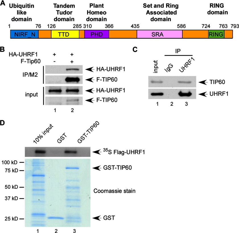
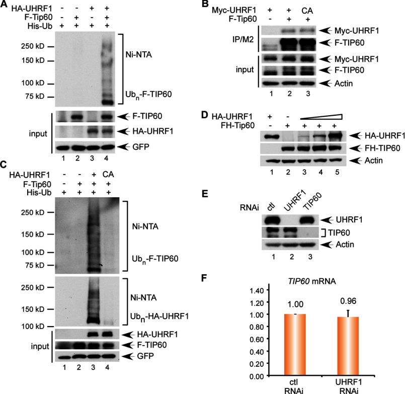
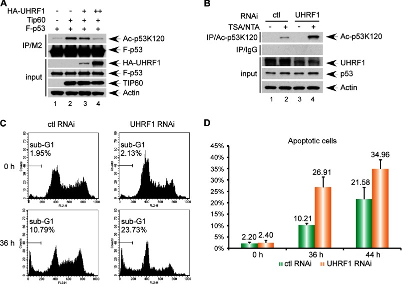
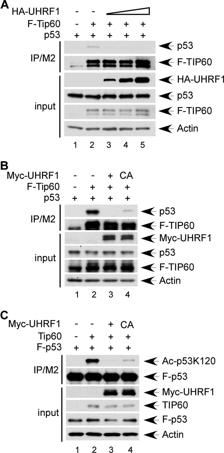
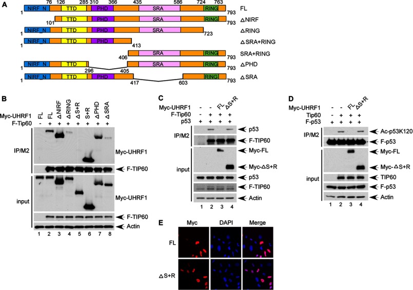
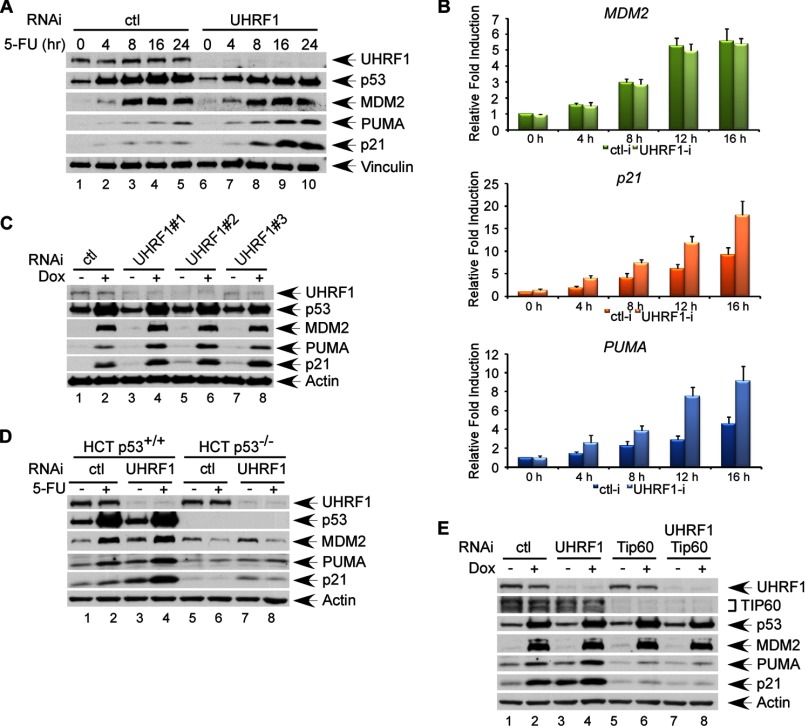
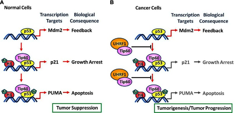
Numerous studies indicate the importance of acetylation in p53-mediated stress responses upon DNA damage. We and others previously showed that TIP60 (Tat-interacting protein of 60 kDa)-mediated acetylation of p53 at K120 is crucial for p53-dependent apoptotic responses. Nevertheless, it remains unclear how TIP60-mediated effects on p53 are dynamically regulated in vivo. Here, we report that UHRF1 (ubiquitin-like with PHD and RING finger domains 1) interacts with TIP60 both in vitro and in vivo and induces degradation-independent ubiquitination of TIP60. Moreover, UHRF1 expression markedly suppresses the ability of TIP60 to acetylate p53. In contrast, RNAi-mediated knockdown of UHRF1 increases the endogenous levels of p53 acetylation at K120 and p53-mediated apoptosis is significantly enhanced in UHRF1-depleted cells. To elucidate the mechanisms of this regulation, we found that the interaction between TIP60 and p53 is severely inhibited in the presence of UHRF1, suggesting that UHRF1 modulates TIP60-mediated functions in both K120 acetylation-dependent and -independent manners. Consistent with this notion, UHRF1 knockdown promotes activation of p21 and PUMA but not MDM2. These findings demonstrate that UHRF1 is a critical negative regulator of TIP60 and suggest that UHRF1-mediated effects on p53 may contribute, at least in part, to its role in tumorigenesis.

Keywords: Acetylation; Apoptosis; Growth Arrest; Oncogene; TIP60; Tumor Suppressor Gene; UHRF1; Ubiquitination; p53.


Figures

FIGURE 1.
FIGURE 1.
UHRF1 is a bona fide TIP60-interacting protein. A, schematic representation of UHRF1 domain structure. B, UHRF1 co-immunoprecipitates with TIP60 in an overexpression system. Whole cell extracts or immunoprecipitates with M2/FLAG antibody from H1299 cells transiently transfected with plasmid DNA expressing HA-UHRF1 or/and FLAG Tip60 were subjected to Western blot with α-FLAG and α-HA antibodies. C, TIP60 interacts with UHRF1 endogenously in U2OS cells. U2OS-derived nuclear extracts or immunoprecipitates with a control IgG or α-UHRF1 antibody were subjected to Western blot with α- TIP60 and α-UHRF1 antibodies. D, UHRF1 interacts with TIP60 directly in vitro. In vitro translated [35S]methionine-labeled 3×FLAG-UHRF1 protein was incubated with purified GST-TIP60 or GST alone. Complexes immobilized with GST resins and recovered using reduced glutathione were subjected to SDS-PAGE and analyzed by autoradiography. The levels of purified GST-TIP60 and GST are shown in the bottom panel stained by Coomassie Blue.
FIGURE 2.
FIGURE 2.
UHRF1 promotes degradation-independent ubiquitination of TIP60. A, UHRF1 induces ubiquitination of TIP60 in vivo. H1299 cells were cotransfected with expression vectors encoding FLAG-Tip60 or/and HA-UHRF1 in combination with His6-ubiquitin. Whole cell extracts and Ni-NTA affinity-purified fractions were analyzed by Western blot with α-FLAG and α-HA antibodies. GFP was used as a control to confirm equal transfection. B, C724A UHRF1 mutant retains interaction with TIP60. H1299 were transiently transfected with expression vectors for FLAG-Tip60 in combination with Myc-tagged wild-type UHRF1 or C724A UHRF1. Whole cell extracts or immunoprecipitates with M2/FLAG antibody were analyzed by Western blot with α-Myc and α-FLAG antibodies. C, UHRF1 directly ubiquitinates TIP60 through its E3 ubiquitin ligase activity. H1299 cells were cotransfected with expression vectors encoding FLAG-Tip60 and His6-ubiquitin alone or together with either HA-UHRF1 or HA-C724A UHRF1 expression vectors. Whole cell extracts and Ni-NTA affinity-purified fractions were analyzed by Western blot with α-FLAG and α-HA antibodies. GFP was used as a control to confirm equal transfection. D, UHRF1 expression does not induce TIP60 degradation. H1299 cells were transfected with FLAG-HA-Tip60 in combination with increasing levels of HA-UHRF1. Whole cell extracts were analyzed by Western blot with α-HA antibody. E, UHRF1 depletion does not affect TIP60 protein level. HCT116 cells were treated with 2 rounds of knock-down with either control RNAi, UHRF1 RNAi, or TIP60 RNAi. Whole cell extracts were analyzed by Western blot with α-UHRF1 and α- TIP60 antibodies. F, UHRF1 depletion does not affect TIP60 mRNA level. Total RNA was extracted from control RNAi or UHRF1 RNAi-treated HCT116. Following reverse transcription, the abundance of TIP60 mRNA was assessed using quantitative real time PCR.
FIGURE 3.
FIGURE 3.
UHRF1 suppresses TIP60-mediated p53 acetylation at K120 and UHRF1 depletion augments damage-induced apoptosis. A, UHRF1 expression inhibits p53 acetylation by TIP60 at K120. H1299 cells were transiently transfected with plasmid DNA expressing FLAG-p53, Tip60, and HA-UHRF1. Total cell extracts and M2 immunoprecipitates were assayed by Western blot using antibodies against HA, p53, and p53-AcK120. B, UHRF1 inactivation significantly increases p53 acetylation at K120. U2OS cells were transiently transfected with either control siRNA or UHRF1 siRNA, and treated for 6 h with 1 μm trichostatin A (TSA) and 5 mm nicotinamde (NTA) prior to harvesting. Cell extracts and immunoprecipitates obtained with α-Acp53K120 or control IgG were analyzed with Western blot using α-UHRF1, α-p53, and α-Acp53K120 antibodies. C, FACS analysis of UHRF1-inactivated U2OS cells treated with etoposide. U2OS cells transiently transfected with either control siRNA or UHRF1 siRNA were treated with 20 μm etoposide for the indicated time. Cells were subsequently fixed in 80% cold methanol, stained with propidium iodide and subjected to DNA content analysis by flow cytometry. D, UHRF1 RNAi increases apoptosis. Apoptosis was assessed as in C, and percentages of apoptotic cells are presented as average values of three independent experiments. Error bars, ±1 S.D.
FIGURE 4.
FIGURE 4.
UHRF1 suppresses TIP60-p53 interaction partially through promoting TIP60 ubiquitination. A, UHRF1 expression inhibits TIP60 interaction with p53. H1299 cells were transiently transfected with expression vectors for FLAG-Tip60 and p53, in the absence of or in combination with HA-UHRF1 expression vector. Cell extracts and M2 immunoprecipitates were assayed by Western blot using α-HA, α-FLAG, and α-p53 antibodies. B, loss of UHRF1-mediated TIP60 ubiquitination partially suppresses TIP60 interaction with p53. H1299 cells were transiently transfected with expression vectors for FLAG-Tip60 and p53 in combination with Myc-tagged UHRF1 or C724A UHRF1. Total cell extracts and M2 immunoprecipitates were assayed by Western blot using antibodies against Myc, FLAG, and p53. C, loss of UHRF1-mediated TIP60 ubiquitination partially inhibits p53 acetylation by TIP60 at K120. H1299 cells were transiently transfected with expression vectors for FLAG-p53, Tip60, and Myc-tagged UHRF1 or C724A UHRF1. Total cell extracts and M2 immunoprecipitates were assayed by Western blot using antibodies against Myc, TIP60, p53, and p53-AcK120.
FIGURE 5.
FIGURE 5.
UHRF1 inhibits TIP60-p53 interaction through its SRA and RING domains. A, schematic representation of UHRF1 deletion mutants used in interaction domain mapping. Full-length UHRF1 and all deletion mutants were subcloned into pCMV-Myc expression vector. B, UHRF1 interacts with TIP60 through its SRA and RING domains. H1299 cells were transiently transfected with expression vectors for FLAG-Tip60 and Myc-tagged UHRF1 deletion constructs. Total cell extracts and M2 immunoprecipitates were assayed by Western blot using antibodies again Myc and FLAG. C, SRA and RING domains of UHRF1 are indispensable for inhibition of TIP60-p53 interaction. H1299 cells were transiently transfected with expression vectors for FLAG-Tip60 and p53, in combination with Myc-tagged full-length UHRF1 or ΔS+R UHRF1. Total cell extracts and M2 immunoprecipitates were analyzed by Western blot using antibodies against Myc, FLAG, and p53. D, UHRF1ΔSRA+RING mutant loses inhibition of TIP60-mediated p53 acetylation at K120. H1299 cells were transiently transfected with expression vectors for FLAG-p53 and Tip60, in combination with Myc-tagged full-length UHRF1 or ΔS+R UHRF1. Total cell extracts and M2 immunoprecipitates were analyzed by Western blot using α-Myc, α- TIP60, α-p53, and α-Acp53K120 antibodies. E, UHRF1ΔSRA+RING mutant retains nuclear localization. U2OS cells were transiently transfected with expression vectors for Myc-tagged full-length UHRF1 or ΔS+R UHRF1. 24 h post-transfection, cells were fixed with paraformaldehyde, immunostained with α-Myc antibody, and counterstained with 4,6-diamidino-2-phenylindole (DAPI). Subcellular localization was assessed by fluorescent microscopy.
FIGURE 6.
FIGURE 6.
UHRF1 depletion up-regulates activation of PUMA and p21 but not MDM2 following DNA damage. A, UHRF1 RNAi in HCT116 cells up-regulates 5-FU induced PUMA and p21 activation but not MDM2 activation. HCT1116 cells were treated with 2 rounds of knock-down with either control RNAi or UHRF1 RNAi. Following treatment with 400 μm 5-FU for the indicated time, whole cell extracts were analyzed by Western blot with the indicated antibodies. B, UHRF1 RNAi in HCT116 cells up-regulates 5-FU induced PUMA and p21 but not MDM2 transcription. HCT116 were treated with 400 μm 5-FU for the indicated time following control RNAi or UHRF1 RNAi. Total RNA was extracted, and cDNA was prepared by reverse transcription. mRNA abundance for MDM2, p21, and PUMA was assessed using quantitative real time PCR. C, UHRF1 RNAi up-regulates doxorubicin induced PUMA and p21 but not MDM2 activation in U2OS cells. U2OS cells were transiently transfected with control siRNA or 3 U2OS-specifc siRNA oligos and treated with or without 0.5 μm doxorubicin for 16 h. Total cell extracts were analyzed by Western blot using the indicated antibodies. D, UHRF1 modulation of damage-induced PUMA and p21 activation is dependent on p53. UHRF1 is inactivated by RNAi in either p53+/+ or p53−/− HCT116 cells. Subsequently, cells were treated with or without 400 μm 5-FU for 8 h before extraction and Western blot analysis using the indicated antibodies. E, UHRF1 modulates damage-induced PUMA and p21 activation in a TIP60-dependent manner. UHRF1 or TIP60 alone, or both UHRF1 and TIP60 were inactivated in U2OS using RNAi. Subsequently, cells were subjected to 16 h of 0.5 μm doxorubicin treatment before extraction and Western blot analysis using the indicated antibodies.
FIGURE 7.
FIGURE 7.
A model for tumorigenesis/tumor progression in cells with UHRF1 overexpression. See text for details.

Similar articles

Cited by

References

    1. Vousden K. H., Prives C. (2009) Blinded by the Light: The Growing Complexity of p53. Cell 137, 413–431 - PubMed
    1. Kruse J. P., Gu W. (2009) Modes of p53 regulation. Cell 137, 609–622 - PMC - PubMed
    1. Dai C., Gu W. (2010) p53 post-translational modification: deregulated in tumorigenesis. Trends Mol. Med. 16, 528–536 - PMC - PubMed
    1. Tang Y., Zhao W., Chen Y., Zhao Y., Gu W. (2008) Acetylation is indispensable for p53 activation. Cell 133, 612–626 - PMC - PubMed
    1. Tang Y., Luo J., Zhang W., Gu W. (2006) Tip60-dependent acetylation of p53 modulates the decision between cell-cycle arrest and apoptosis. Mol. Cell 24, 827–839 - PubMed

Publication types

MeSH terms

LinkOut - more resources