. 2013 Jun 28;288(26):19116-26.
doi: 10.1074/jbc.M113.469007. Epub 2013 May 15.

HIV-2 and SIVmac accessory virulence factor Vpx down-regulates SAMHD1 enzyme catalysis prior to proteasome-dependent degradation

Affiliations

HIV-2 and SIVmac accessory virulence factor Vpx down-regulates SAMHD1 enzyme catalysis prior to proteasome-dependent degradation

Maria DeLucia et al. J Biol Chem. .

Abstract

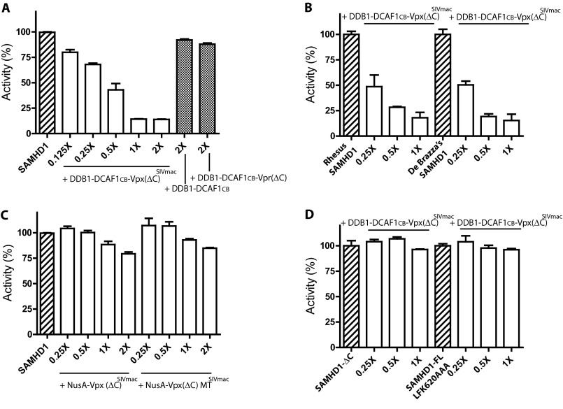
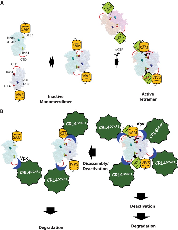
SAMHD1, a dGTP-regulated deoxyribonucleoside triphosphate (dNTP) triphosphohydrolase, down-regulates dNTP pools in terminally differentiated and quiescent cells, thereby inhibiting HIV-1 infection at the reverse transcription step. HIV-2 and simian immunodeficiency virus (SIV) counteract this restriction via a virion-associated virulence accessory factor, Vpx (Vpr in some SIVs), which loads SAMHD1 onto CRL4-DCAF1 E3 ubiquitin ligase for polyubiquitination, programming it for proteasome-dependent degradation. However, the detailed molecular mechanisms of SAMHD1 recruitment to the E3 ligase have not been defined. Further, whether divergent, orthologous Vpx proteins, encoded by distinct HIV/SIV strains, bind SAMHD1 in a similar manner, at a molecular level, is not known. We applied surface plasmon resonance analysis to assess the requirements for and kinetics of binding between various primate SAMHD1 proteins and Vpx proteins from SIV or HIV-2 strains. Our data indicate that Vpx proteins, bound to DCAF1, interface with the C terminus of primate SAMHD1 proteins with nanomolar affinity, manifested by rapid association and slow dissociation. Further, we provide evidence that Vpx binding to SAMHD1 inhibits its catalytic activity and induces disassembly of a dGTP-dependent oligomer. Our studies reveal a previously unrecognized biochemical mechanism of Vpx-mediated SAMHD1 inhibition: direct down-modulation of its catalytic activity, mediated by the same binding event that leads to SAMHD1 recruitment to the E3 ubiquitin ligase for proteasome-dependent degradation.

Keywords: Enzyme Turnover; Enzymes; HIV; Restriction Factors; Ubiquitin Ligase; Ubiquitination; Virulence Factors.


Figures

FIGURE 1.
FIGURE 1.
Schematic representation of human SAMHD1 and alignment of Vpx protein sequences. A, SAMHD1 comprises two folded domains: a SAM (residues 42–110) and a dNTPase domain (residues 128–576). The dNTPase domain encompasses a metal-dependent phosphohydrolase homologous region with a conserved HD motif (residues 160–325). Several residues that have been characterized for their roles in SAMHD1 function are indicated. The nuclear localization signal is at residues 11–14 (14, 15). Residues Asp-137, Gln-142, Arg-145, and Arg-451 are located at the dGTP-binding allosteric site, and residues Arg-164, His-206, Asp-207, and Asp-311 are at the catalytic site (8). The C-terminal residues Arg-617, Leu-620, Phe-621, and Met-626 are recognized by Vpx for proteasome-dependent down-regulation (20, 27). B, alignment of Vpx amino acid sequences encoded by SIVmac, HIV-2 Rod9, and HIV-2 7312a. The residues critical for SAMHD1 recruitment and down-regulation (20) are indicated in the box.
FIGURE 2.
FIGURE 2.
Real-time kinetic analysis of SAMHD1 and DDB1-DCAF1-Vpx interactions. A, SPR sensorgrams of DDB1-DCAF1CB-Vpx(ΔC)SIVmac binding to GST-Hu SAMHD1-FL (A), and GST-rhesus (GST-Rh) SAMHD1-FL (B). The concentrations of DDB1-DCAF1CB-Vpx(ΔC)SIVmac were 8, 16, 32, 64, 128, 256, 512, 1024, and 2048 nm. The average association rates for two independent experiments are summarized in Table 1. C, binding curves of DDB1-DCAF1CB-Vpx(ΔC)SIVmac to human (Hu; solid circles), rhesus (Rh; solid squares), and De Brazza's (DB; solid triangles) SAMHD1 were generated by plotting the response levels (RU) at 120 s (marked as dissociation in A and B). The average Kd values for the interactions between human, rhesus, or De Brazza's SAMHD1 protein and the DDB1-DCAF1CB-Vpx(ΔC)SIVmac complex, for two independent experiments, are summarized in Table 1. D, SPR sensorgrams of DDB1-DCAF1CB-Vpx(ΔC)SIVmac binding to GST-Hu SAMHD1-CTD (residues 595–626). E, SPR sensorgrams of DDB1-DCAF1CB-Vpx(ΔC)SIVmac, with Ala substitution at Glu-15 of Vpx (E15A), binding to GST-Hu SAMHD1-CTD. F, binding curve of DDB1-DCAF1CB-Vpx(ΔC)SIVmac to human SAMHD1-CTD is shown. The data were analyzed as described in C. The average kon and Kd values are shown in Table 1. Response levels (RU) of DDB1-DCAF1CB-Vpx(ΔC)SIVmac, with Vpx alanine substitutions at Asn-12, Glu-15, Glu-16, or Thr-17 binding to human SAMHD1-CTD are also shown.
FIGURE 3.
FIGURE 3.
Both the C terminus of SAMHD1 and the N-terminal region of Vpx are essential for proteasome-dependent degradation of SAMHD1. A, in vitro ubiquitination assays of WT or C-terminally deleted (-ΔC) Human, De Brazza's, and rhesus SAMHD1 with CRL4-DCAF1CB-Vpx7312a. Control reactions were performed with DDB1-DCAF1CB, without Vpx, and all other CRL4 components with or without WT SAMHD1. B, HEK293 cells were transiently co-transfected with full-length DCAF1 (DCAF1-FL), SIVmac Vpx, and various human SAMHD1 constructs (SAMHD1-FL, WT; SAMHD1-ΔC, residues 1–595; ΔN-SAMHD1, residues 113–626) as indicated. The levels of ectopically expressed proteins were determined by immunoblotting with appropriate antibodies after separating cell lysates by SDS-PAGE, 48 h after transfection. Each transfection condition was carried out twice. C, HEK293 cells were transiently co-transfected with DCAF1CA, human (Hu) SAMHD1, and SIVmac Vpx and analyzed as in B after cells were treated with MG132 (+) or mock-treated (−). D, HEK293 cells were transiently co-transfected with human SAMHD1-FL, DCAF1CA (residues 1040–1400), and VpxSIVmac, VpxRod9, or Vpx7312a WT or mutant (E15A/E16A for SIVmac and HIV-2 Rod9; E14A/E15A for HIV-2 7312a), as indicated. Protein levels were determined as described in B. E, human SAMHD1 proteins were co-expressed with DCAF1CA and Vpx7312a WT or two mutants (D13G or E14A/E15A) and analyzed as in B.
FIGURE 4.
FIGURE 4.
SAMHD1 forms catalytically active dGTP-dependent tetramers. A, quaternary states of human and six monkey SAMHD1 proteins were characterized by chemical cross-linking assays. B, dGTP-dependent tetramer formation of rhesus and De Brazza's SAMHD1 proteins was assessed by analytical size exclusion column chromatography. The peak elution volumes are indicated. C, the relative dNTPase activities of human and monkey SAMHD1. Error bars, S.D.
FIGURE 5.
FIGURE 5.
The recruitment of SAMHD1 to DDB1-DCAF1-Vpx via its C terminus inhibits the dNTPase catalytic activity. A, the dNTPase activity of human SAMHD1 was determined in the presence of increasing concentrations of DDB1-DCAF1CB-Vpx(ΔC)SIVmac (molar ratio ranging from 0.125 to 2). The relative dNTPase activity toward dATP in each reaction was calculated and normalized over the catalytic activity of SAMHD1 alone (hashed bar). The catalytic activity of SAMHD1 in the presence of DDB1-DCAF1CB or DDB1-DCAF1CB-Vpr(ΔC) (residues 1–79) was also determined (solid bars). B, the relative dNTPase activity of rhesus and De Brazza's SAMHD1 was measured with increasing concentrations of DDB1-DCAF1CB-Vpx(ΔC)SIVmac at molar ratios ranging from 0.25 to 2. C, the catalytic activity of SAMHD1 was determined with increasing concentrations of NusA-VpxSIVmac WT and mutant (N12A/E15A/E16A/T17A) at molar ratios of 0.25–2-fold. D, the relative dNTPase activity of SAMHD1-ΔC or SAMHD1-FL with a triple residue mutation (L620A/F621A/K622A) was determined with increasing concentrations of DDB1-DCAF1c-Vpx(ΔC)SIVmac, as indicated. Error bars, S.D.
FIGURE 6.
FIGURE 6.
DDB1-DCAF1-Vpx breaks dGTP-SAMHD1 tetramers into dimeric and monomeric DDB1-DCAF1-Vpx-SAMHD1 supramolecular complexes. A, multiangle light scattering of SAMHD1, DDB1-DCAF1-VpxSIVmac, and their mixtures. SAMHD1 alone (green trace) or its mixture with DDB1-DCAF1CB-Vpx(ΔC)SIVmac (red trace) was injected into an analytical gel filtration column at a flow rate of 0.5 ml/min, and the absorbance trails were recorded at 280 nm. The estimated molecular mass of protein complexes in the peaks is shown across the elution peaks using the same color scheme. B, SAMHD1 preincubated with dGTP (4 mm) (green trace) and its mixture with increasing concentrations of DDB1-DCAF1CB-Vpx(ΔC)SIVmac (red and blue traces) were separated over an analytical gel filtration column (top). Data were analyzed as described in A. The elution fractions (every 0.25 ml) from the gel filtration analysis of the SAMHD1, dGTP, and high concentration of DDB1-DCAF1-Vpx(ΔC)SIVmac mixture (blue trace) were collected, concentrated, separated over SDS-PAGE, and visualized by Coomassie Blue staining. Lanes 3, 7, 11, 15, and 19 are labeled with the corresponding elution volumes from the gel filtration column chromatography at the top.
FIGURE 7.
FIGURE 7.
Model of SAMHD1 activation by dGTP and recruitment to CRL4-DCAF1 reprogrammed by Vpx. A, dGTP-induced SAMHD1 tetramerization and activation. Binding of dGTP (black in the tetramer) to each allosteric site of four SAMHD1 monomers induces tetramerization via a transient head-to-tail dimer. Residues near the allosteric sites are shown in different colors for each monomer to help with orientation: Asp-137 (head) and Arg-451 (tail). The catalytic site is indicated with His-206 (H206) and Asp-207 (D207). The structural model of the HD domain was generated based on the crystal structures of EF1143 (Protein Data Bank code 3IRH) (44) and the HD domain of SAMHD1 (3UN1) (8) and was rendered faintly to emphasize residues and dGTP molecules. B, SAMHD1 monomer, dimer, and tetramer are recruited to CRL4-DCAF1 by Vpx. Vpx, while binding to DCAF1 via its middle region (16), interacts with the C terminus of SAMHD1 via its N terminus. DDB1-DCAF1-Vpx binding to the SAMHD1 tetramer deactivates its dNTPase activity and subsequently disassembles it to dimers and monomers and induces proteasome-dependent degradation.

Similar articles

Cited by

References

    1. Berger A., Sommer A. F., Zwarg J., Hamdorf M., Welzel K., Esly N., Panitz S., Reuter A., Ramos I., Jatiani A., Mulder L. C., Fernandez-Sesma A., Rutsch F., Simon V., König R., Flory E. (2011) SAMHD1-deficient CD14+ cells from individuals with Aicardi-Goutieres syndrome are highly susceptible to HIV-1 infection. PLoS Pathog. 7, e1002425. - PMC - PubMed
    1. Hrecka K., Hao C., Gierszewska M., Swanson S. K., Kesik-Brodacka M., Srivastava S., Florens L., Washburn M. P., Skowronski J. (2011) Vpx relieves inhibition of HIV-1 infection of macrophages mediated by the SAMHD1 protein. Nature 474, 658–661 - PMC - PubMed
    1. Laguette N., Sobhian B., Casartelli N., Ringeard M., Chable-Bessia C., Ségéral E., Yatim A., Emiliani S., Schwartz O., Benkirane M. (2011) SAMHD1 is the dendritic- and myeloid-cell-specific HIV-1 restriction factor counteracted by Vpx. Nature 474, 654–657 - PMC - PubMed
    1. Descours B., Cribier A., Chable-Bessia C., Ayinde D., Rice G., Crow Y., Yatim A., Schwartz O., Laguette N., Benkirane M. (2012) SAMHD1 restricts HIV-1 reverse transcription in quiescent CD4+ T-cells. Retrovirology 9, 87. - PMC - PubMed
    1. Baldauf H. M., Pan X., Erikson E., Schmidt S., Daddacha W., Burggraf M., Schenkova K., Ambiel I., Wabnitz G., Gramberg T., Panitz S., Flory E., Landau N. R., Sertel S., Rutsch F., Lasitschka F., Kim B., König R., Fackler O. T., Keppler O. T. (2012) SAMHD1 restricts HIV-1 infection in resting CD4+ T cells. Nat. Med. 18, 1682–1687 - PMC - PubMed

Publication types

MeSH terms

Substances

LinkOut - more resources