. 2013 May 15;33(20):8596-607.
doi: 10.1523/JNEUROSCI.2083-12.2013.

Shh/Boc signaling is required for sustained generation of ipsilateral projecting ganglion cells in the mouse retina

Affiliations

Shh/Boc signaling is required for sustained generation of ipsilateral projecting ganglion cells in the mouse retina

Luisa Sánchez-Arrones et al. J Neurosci. .

Abstract

Sonic Hedgehog (Shh) signaling is an important determinant of vertebrate retinal ganglion cell (RGC) development. In mice, there are two major RGC populations: (1) the Islet2-expressing contralateral projecting (c)RGCs, which both produce and respond to Shh; and (2) the Zic2-expressing ipsilateral projecting RGCs (iRGCs), which lack Shh expression. In contrast to cRGCs, iRGCs, which are generated in the ventrotemporal crescent (VTC) of the retina, specifically express Boc, a cell adhesion molecule that acts as a high-affinity receptor for Shh. In Boc(-/-) mutant mice, the ipsilateral projection is significantly decreased. Here, we demonstrate that this phenotype results, at least in part, from the misspecification of a proportion of iRGCs. In Boc(-/-) VTC, the number of Zic2-positive RGCs is reduced, whereas more Islet2/Shh-positive RGCs are observed, a phenotype also detected in Zic2 and Foxd1 null embryos. Consistent with this observation, organization of retinal projections at the dorsal lateral geniculate nucleus is altered in Boc(-/-) mice. Analyses of the molecular and cellular consequences of introducing Shh into the developing VTC and Zic2 and Boc into the central retina indicate that Boc expression alone is insufficient to fully activate the ipsilateral program and that Zic2 regulates Shh expression. Taking these data together, we propose that expression of Boc in cells from the VTC is required to sustain Zic2 expression, likely by regulating the levels of Shh signaling from the nearby cRGCs. Zic2, in turn, directly or indirectly, counteracts Shh and Islet2 expression in the VTC and activates the ipsilateral program.


Figures

Figure 1.
Figure 1.
Boc−/− mice present a reduction of uncrossed retinal projections. A–H, Ventral views of the intact brains (A–D) and frontal (E–F′) or horizontal (G,H) vibratome sections through the contralateral and ipsilateral dLGN (E–F′) and optic chiasm (G,H) from E18 and P4 WT and Boc−/− animals (as indicated in the panels) with unilateral DiI filling of the optic disk to visualize visual fiber trajectory. There is strong reduction of projecting fibers in the ipsilateral lateral geniculate nucleus (iLGN) of the mutants. *Ipsilateral reduction. F, Arrowheads indicate growth cones in the initial portion of the mutant ipsilateral optic tract. I, The graph compares the values of the ipsilateral index in WT and Boc−/− mice at embryonic and postnatal stages. The number of analyzed animals is indicated in the graph for each stage. *p < 0.05 (Student's unpaired t test). **p < 0.01 (Student's unpaired t test). cLGN, Contralateral lateral geniculate nucleus; cot, contralateral optic chiasm; iot, ipsilateral optic tract. Scale bars: A–D, 1000 μm; E, F, 500 μm.
Figure 2.
Figure 2.
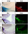
Boc expression is downregulated in the VTC of Zic2kd/kd embryos. Frontal cryostat sections from E14.5 Boc+/lacZ and 16.5 WT and Zic2kd/kd central retinas reacted with with X-gal histochemistry, hybridized with probes for Boc or immunostained with antibodies against Zic2 and the related Cdon. A, B, The specific expression of Boc in the VTC is particularly abundant in the Zic2-positive domain. C–F, Boc expression is absent in Zic2kd/kd embryos but maintained in the CMZ where Cdon is also localized. cmz, Ciliary marginal zone; L, lens. Scale bar, 200 μm.
Figure 3.
Figure 3.
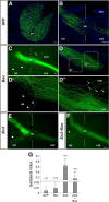
Boc is insufficient to activate the ipsilateral program. A–F, Confocal images of flat-mounted electroporated retina (A) and representative sections of the optic chiasm (B–F) from E18 embryos electroporated at E13 with a plasmid expressing GFP alone (A,B), or GFP with Boc (C,D), Zic2 (E), or Zic2 and Boc (F). Electroporations mostly targeted the central retina (A). D′, D″, High-magnification views of the areas boxed in D. In embryos electroporated with Boc, most axons (C,D) cross the midline (dotted lines), whereas a few axons grow into the ipsilateral optic tract (D, D′, arrowheads) and in larger numbers in the contralateral optic nerve (D″, arrows). G, The graph compares the values of the ipsilateral index in embryos electroporated with the different constructs. The number of analyzed animals is indicated on top of each bar. ***p < 0.001, compared with GFP (Student's unpaired t test). od, Optic disc; con, contralateral optic nerve; oc, optic chiasm; iot, ipsilateral optic tract; cot, contralateral optic tract. Scale bar, 100 μm.
Figure 4.
Figure 4.
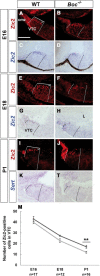
A–L, Boc is required to maintain the appropriate number of Zic2-positive iRGCs. Frontal cryostat sections from E16 (A–D), E18 (E–H), and P1 (I–L) WT and Boc−/− central retinas immunostained for Zic2 or hybridized with specific probes for Zic2 and Sert as indicated in the panels. The loss of Zic2 immunostaining is preceded by a decrease in its mRNA expression (compare C and G with D and H). There is a progressive decrease and distal clustering of Zic2/Sert-positive and iRGCs in the Boc−/− VTC. M, The graph represents the percentage of Zic2+ cells in the VTC of WT (dark gray line) and mutant (light gray line) animals at different stages. **p < 0.01 (Student's unpaired t test). cmz, Ciliary marginal zone; L, lens. Scale bar, 200 μm.
Figure 5.
Figure 5.
A–L′, Boc is required to restrain Islet2/Shh expression within the VTC. Frontal cryostat sections from E16 (A–D′), E18 (E–H′), and P0 (I–L′) WT and Boc−/− central retinas immunostained for Brn3b, double-stained for Zic2 (green) and Islet2 (red), or hybridized with specific probes for Isl2 and Shh, as indicated in the panels. The increase in Islet2+ cells parallels the decrease in Zic2+ cells in Boc−/− tissue. There is a correspondent increase in Islet2 and Shh expression in the mutants starting from E18. Dashed lines delimit the VTC. M–O′, Low-power views of flat-mounted preparations of the ipsilateral and contralateral retinas from E17 embryos subjected to unilateral retrograde labeling from the optic tract, double-labeled with antibodies against Zic2 (O,O′). In WT, the majority of backfilled neurons localized to the ipsilateral VTC (the VTC area is marked with dotted lines), whereas only few RGCs were found in the contralateral VTC. In Boc−/− VTC, this proportion is shifted and there are numerous Zic2+ backfilled cells in the cVTC (O′). P, Q, The histograms represent the amount of Zic2+, Islet2+, or Brn3b+ cells in P0 (P) and the amount of backfilled cells present in the ipsilateral and contralateral retinas of E17 (Q), and WT (dark gray bars) and Boc−/− (light gray bars) VTC. **p < 0.01 (Student's unpaired t test). ***p < 0.001 (Student's unpaired t test). cmz, Ciliary marginal zone. Scale bar, 100 μm.
Figure 6.
Figure 6.
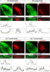
Organization of the retino-geniculate projections in postnatal Boc−/− mice. Confocal analysis of frontal sections through the dLGN of P12 and P18 WT and Boc−/− mice (as indicated in the panels) after whole-eye anterograde tracing with green and red fluorescent dyes. C′, F′, I′, L′, Reslice profiles taken at the levels indicated in C, F, I, and L, respectively. The accompanying graphs represent the spectra of red (ipsilateral) and green (contralateral) channels of reslice 2 (C′,F′) and reslice 3 (I′,L′), respectively. In WT, the ipsilateral projection (red) is clustered in the dorsocentral dLGN surrounded by contralateral terminations (green). In Boc−/−, ipsilateral fibers are reduced in this region. There is increased clustering of fiber in the dorsomedial tip (area circled with dotted lines) of dLGN where late-born cRGCs of the VTC project. d, Dorsal; m, medial; l, lateral; v, ventral. Scale bar: A–L, 250 μm.
Figure 7.
Figure 7.
Organization of the retino-geniculate projections in adult Boc−/− mice. The images show confocal analysis of frontal sections through different axial levels of the dLGN of P30 WT and Boc−/− mice after whole-eye anterograde tracing with green and red fluorescent dyes. Reslice profiles represented below each image are taken at the levels indicated in A for A–D, and in E for E–H. The accompanying graphs represent the spectra of red (ipsilateral) and green (contralateral) channels of the reslices as indicated in the figure. d, Dorsal; m, medial; l, lateral; v, ventral. Scale bar, 250 μm.
Figure 8.
Figure 8.
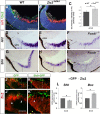
A feedback regulatory loop between Zic2 and Shh signaling in the VTC. A, B, D, E–I, Frontal cryostat sections from E16 WT (A, D, G), Zic2kd/kd (B, E, H), and Foxd1−/− (F, I) central retinal sections hybridized with probes for Islet2 (A, B, D–F) and Shh (G–I) and immunostained with antibodies against Zic2 (A,B). WT embryos are Zic2kd/kd littermates. Littermates from Foxd1−/− line are not shown as they were no different from those shown in D and G. C, The histogram represents the increment of Islet2+ fluorescence in the Zic2kd/kd VTC compared with WT. *p < 0.05 (Student's unpaired t test). J–K′, Control GFP (J, J′) or Shh::GFP (K, K′) electroporated VTC from E15 WT embryos immunostained with antibodies against Zic2 (red). There is an absence of Zic2 staining in the region where Shh is ectopically expressed, whereas Zic2 staining is still present in GFP-positive controls (solid and empty arrowheads in J–K′ point to Zic2+ and − regions, respectively). L, qPCR analysis of the levels of Shh and Boc expression after ex utero electroporation of Zic2 in the central retina of E13 WT embryos. Tissue was collected and analyzed 24 h later. Analysis was performed on pools of eight different retinas for each sample, and experiments were repeated three times. Data were analyzed with the Mann–Whitney test: *p < 0.05. cmz, Ciliary marginal zone; L, lens. Scale bars: A–I, 200 μm; J–K′, 100 μm.
Figure 9.
Figure 9.
Summary diagram of the proposed mechanism for iRGC and cRGC specification in the VTC and its consequent retinotopic organization based on the analysis of Boc-null mice. A, A feedback regulatory loop between Boc-mediated Shh signaling and Zic2 activated by low Shh concentration drives iRGC generation in WT animals. In the absence of Boc, the VTC receives higher Shh levels from the adjacent cRGC precluding Zic2 expression and thus shifting RGC toward a contralateral identity (see Discussion). B, Shift of RGC identity coupled to occasional pathfinding defects causes changes in the organization of retinal projections at the dLGN. cmz, Ciliary marginal zone; L, lens.

Similar articles

Cited by

References

    1. Allen BL, Song JY, Izzi L, Althaus IW, Kang JS, Charron F, Krauss RS, McMahon AP. Overlapping roles and collective requirement for the coreceptors GAS1, CDO, and BOC in SHH pathway function. Dev Cell. 2011;20:775–787. doi: 10.1016/j.devcel.2011.04.018. - DOI - PMC - PubMed
    1. Aruga J. The role of Zic genes in neural development. Mol Cell Neurosci. 2004;26:205–221. doi: 10.1016/j.mcn.2004.01.004. - DOI - PubMed
    1. Aruga J, Tohmonda T, Homma S, Mikoshiba K. Zic1 promotes the expansion of dorsal neural progenitors in spinal cord by inhibiting neuronal differentiation. Dev Biol. 2002;244:329–341. doi: 10.1006/dbio.2002.0598. - DOI - PubMed
    1. Bae GU, Domené S, Roessler E, Schachter K, Kang JS, Muenke M, Krauss RS. Mutations in CDON, encoding a hedgehog receptor, result in holoprosencephaly and defective interactions with other hedgehog receptors. Am J Hum Genet. 2011;89:231–240. doi: 10.1016/j.ajhg.2011.07.001. - DOI - PMC - PubMed
    1. Balaskas N, Ribeiro A, Panovska J, Dessaud E, Sasai N, Page KM, Briscoe J, Ribes V. Gene regulatory logic for reading the Sonic Hedgehog signaling gradient in the vertebrate neural tube. Cell. 2012;148:273–284. doi: 10.1016/j.cell.2011.10.047. - DOI - PMC - PubMed

Publication types

MeSH terms