. 2013 Jul;89(1):123-34.
doi: 10.1111/mmi.12263. Epub 2013 Jun 7.

Superoxide poisons mononuclear iron enzymes by causing mismetallation

Affiliations

Superoxide poisons mononuclear iron enzymes by causing mismetallation

Mianzhi Gu et al. Mol Microbiol. 2013 Jul.

Abstract

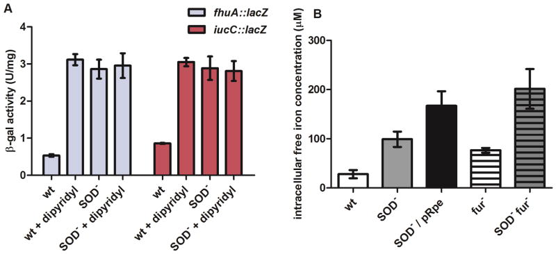
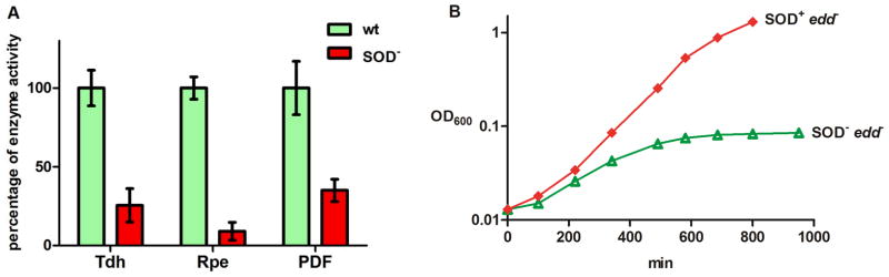
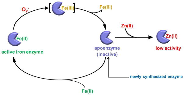
Superoxide (O(2)(-)) is a primary agent of intracellular oxidative stress. Genetic studies in many organisms have confirmed that excess O(2)(-) disrupts metabolism, but to date only a small family of [4Fe-4S] dehydratases have been identified as direct targets. This investigation reveals that in Escherichia coli O(2)(-) also poisons a broader cohort of non-redox enzymes that employ ferrous iron atoms as catalytic cofactors. These enzymes were inactivated by O(2)(-) both in vitro and in vivo. Although the enzymes are known targets of hydrogen peroxide, the outcome with O(2)(-) differs substantially. When purified enzymes were damaged by O(2)(-) in vitro, activity could be completely restored by iron addition, indicating that the O(2)(-) treatment generated an apoprotein without damaging the protein polypeptide. Superoxide stress inside cells caused the progressive mismetallation of these enzymes with zinc, which confers little activity. When O(2)(-) stress was terminated, cells gradually restored activity by extracting zinc from the proteins. The overloading of cells with zinc caused mismetallation even without O(2)(-) stress. These results support a model in which O(2)(-) repeatedly excises iron from these enzymes, allowing zinc to compete with iron for remetallation of their apoprotein forms. This action substantially expands the physiological imprint of O(2)(-) stress.


Figures

Fig. 1
Fig. 1. Endogenous O2 disrupts Fur metallation
(A) The Fur regulon is derepressed in SOD-deficient strains in defined medium. The wild type (wt) and SOD-deficient (SOD) strains were grown aerobically in glucose/amino acid M9 medium to an OD600 of ~0.25. Where indicated, 1 mM dipyridyl was added, and cells were aerated for an additional 30 min. Supplemental Figure S1 shows that the derepression was mediated by loss of Fur activity. (B) Intracellular free iron levels. Cells were cultured as described in (A). To induce Rpe expression, 0.5 mM IPTG was added to the culture at ~0.1 OD600. EPR samples were prepared as described in “Experimental procedures”. Wild type, AB1157; SOD, PN134; fur, KK210; and SOD fur, KK216.
Fig. 2
Fig. 2. (A) Mononuclear iron enzymes are damaged in the SOD-deficient strains
The wild type (wt) and SOD-deficient (SOD) strains were grown aerobically in glucose/amino acid minimal A medium to an OD600 of ~0.3, and enzyme activities were measured. (B) The pentose phosphate pathway fails in SOD mutants. Cells were precultured anaerobically and then diluted into aerobic gluconate/amino acid minimal A medium. Cell growth was monitored thereafter. The Δedd mutants lack 6-phosphogluconate dehydratase and therefore depend exclusively upon the pentose-phosphate pathway to catabolize gluconate.
Fig. 3
Fig. 3. Superoxide rapidly inactivates mononuclear iron enzymes in vitro
Purified Tdh (A), Rpe (B) and PDF (C) were metallated with Fe(II) anaerobically. O2 was generated in vitro aerobically by xanthine and xanthine oxidase. Where indicated, 500 U/ml of SOD was added before exposure to O2.
Fig. 4
Fig. 4. Addition of Fe2+ fully restores activity to O2-damaged Tdh (A) and Rpe (B)
Iron-charged enzymes were treated with O2 for 3 min in the presence of catalase, and 500 U/ml of SOD was then added to scavenge residual O2. Where indicated, 0.5 mM Fe(NH4)2(SO4)2 was then added to reactivate the enzymes, and 0.5 mM ascorbic acid was also included to keep iron in its ferrous form in the aerobic environment.
Fig. 5
Fig. 5. O2 damages Tdh in vivo by causing mismetallation
(A) Tdh progressively loses activity in the SOD strain. Wild type (wt) and SOD mutant were grown anaerobically to an OD600 of 0.2, de novo protein synthesis was stopped, and cultures were shifted to aerobic conditions. At indicated time points, aliquots were taken to measure Tdh activity. (B) Tdh does not accumulate in the apoprotein form in O2 -stressed cells. Cell extracts were prepared from SOD strain which had been aerated for 120 min. Where indicated, 0.5 mM of Fe(NH4)2(SO4)2 was added to the extracts. (C) Tdh protein polypeptide is not degraded in O2-stressed cells. Cell extracts were prepared from wild type and SOD strain which had been aerated for 120 min. The extracts were incubated anaerobically with 2.5 mM EDTA at RT for 10 min. The enzyme was then charged with Fe(NH4)2(SO4)2 under anaerobic conditions prior to assay (Experimental Procedures). (D) Tdh from O2 -stressed cells is not metallated with iron. The cell extracts from part (C) were treated with 50 μM H2O2 anaerobically.
Fig. 6
Fig. 6. Rpe (A) and Tdh (B) restore activity in vivo after superoxide stress is removed
The SOD deficient strain (SOD) was grown anaerobically at 37 °C to an OD600 of ~0.20. Then de novo protein synthesis was stopped, and cells were shifted to aerobic conditions. After 3 hours of aeration at 37 °C, cells were returned to anaerobic conditions. At the subsequent time points, aliquots were harvested for measurement of Rpe and Tdh activity.
Fig. 7
Fig. 7
Model for in vivo inactivation of mononuclear iron enzymes by O2.
Fig. 8
Fig. 8. Even in the absence of O2 stress, excess intracellular zinc causes Tdh mismetallation in vivo
The wild type strain (wt) and the zinc efflux deficient mutant (ΔzntA) were grown in glucose/amino acid medium supplemented with 150 μM ZnCl2. To test the H2O2 sensitivity of Tdh, 50 μM H2O2 was incubated with the crude extracts at RT for 3 min. To chelate metals from Tdh active site, the crude extracts were anaerobically incubated with 2.5 mM EDTA at RT for 10 min. The enzyme was then charged with Fe(NH4)2(SO4)2 or ZnCl2 prior to assay.

References

    1. Akana J, Fedorov AA, Fedorov E, Novak WR, Babbitt PC, Almo SC, Gerlt JA. D-ribulose 5-phosphate 3-epimerase: functional and structural relationships to members of the ribulose-phosphate binding (β/α) 8-barrel superfamily. Biochemistry. 2006;45:2493–2503. - PubMed
    1. Al-Maghrebi M, Fridovich I, Benov L. Manganese supplementation relieves the phenotypic deficits seen in superoxide-dismutase-null Escherichia coli. Arch Biochem Biophys. 2002;402:104–109. - PubMed
    1. Altuvia S, Almiron M, Huisman G, Kolter R, Storz G. The dps promoter is activated by OxyR during growth and by IHF and sigma S in stationary phase. Mol Microbiol. 1994;13:265–272. - PubMed
    1. Anbar AD. Elements and evolution. Science. 2008;322:1481–1483. - PubMed
    1. Anjem A, Imlay JA. Mononuclear iron enzymes are primary targets of hydrogen peroxide stress. J Biol Chem. 2012;287:15544–15556. - PMC - PubMed

Publication types

LinkOut - more resources