. 2013 May 16:14:333.
doi: 10.1186/1471-2164-14-333.

Genomic and physiological variability within Group II (non-proteolytic) Clostridium botulinum

Affiliations

Genomic and physiological variability within Group II (non-proteolytic) Clostridium botulinum

Sandra C Stringer et al. BMC Genomics. .

Abstract

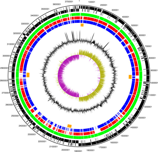
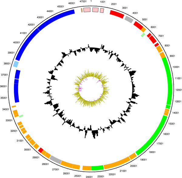
Background: Clostridium botulinum is a group of four physiologically and phylogenetically distinct bacteria that produce botulinum neurotoxin. While studies have characterised variability between strains of Group I (proteolytic) C. botulinum, the genetic and physiological variability and relationships between strains within Group II (non-proteolytic) C. botulinum are not well understood. In this study the genome of Group II strain C. botulinum Eklund 17B (NRP) was sequenced and used to construct a whole genome DNA microarray. This was used in a comparative genomic indexing study to compare the relatedness of 43 strains of Group II C. botulinum (14 type B, 24 type E and 5 type F). These results were compared with characteristics determined from physiological tests.

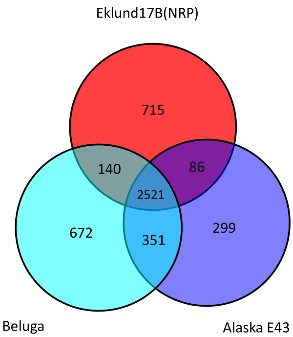
Results: Whole genome indexing showed that strains of Group II C. botulinum isolated from a wide variety of environments over more than 75 years clustered together indicating the genetic background of Group II C. botulinum is stable. Further analysis showed that strains forming type B or type F toxin are closely related with only toxin cluster genes targets being unique to either type. Strains producing type E toxin formed a separate subset. Carbohydrate fermentation tests supported the observation that type B and F strains form a separate subset to type E strains. All the type F strains and most of type B strains produced acid from amylopectin, amylose and glycogen whereas type E strains did not. However, these two subsets did not differ strongly in minimum growth temperature or maximum NaCl concentration for growth. No relationship was found between tellurite resistance and toxin type despite all the tested type B and type F strains carrying tehB, while the sequence was absent or diverged in all type E strains.

Conclusions: Although Group II C. botulinum form a tight genetic group, genomic and physiological analysis indicates there are two distinct subsets within this group. All type B strains and type F strains are in one subset and all type E strains in the other.


Figures

Figure 1
Figure 1
Circular figure showing relatedness of the chromosome of sequenced Group II C. botulinum strains. Concentric rings represent the genomes of Eklund 17B (NRP) and three other sequenced strains of Group II C. botulinum. From outermost to innermost ring: 1. Black: DNA coordinates for Eklund 17B (NRP) DNA sequence 2. Black: Forward and reverse frames coding sequences for Eklund 17B (NRP). 3. Green: Orthologous coding sequences for Eklund 17B (JGI) (forward only). 4. Red: Orthologous coding sequences shared with Alaska E43 (forward only). 5. Blue: Orthologous coding sequences shared with Beluga (forward only). 6. Orange: Predicted intact bacteriophage sequences in Eklund 17B (NRP). 7. Black: GC content plot as a percentage for Eklund 17B (NRP). 8. Purple/green: GC skew plot for Eklund 17B (NRP). The figure indicates the rRNA operons as regions containing no coding sequences and also represented by a clear spike in the GC content plot present near the replication origin and termination regions. The GC skew plot shows a positive skew on the leading strand.
Figure 2
Figure 2
Venn diagram showing the relatedness of Group II C. botulinum strains. Orthologous CDSs were calculated between the three available Group II C. botulinum genome sequences by reciprocal FASTA best match analysis. CDSs shared between specific strains and CDSs unique to a particular strain are plotted on the Venn diagram.
Figure 3
Figure 3
Circular figure showing features of Group II C. botulinum Eklund 17B (NRP) plasmid pCLL. Concentric rings show the following from outermost to innermost. 1–2. Plasmid features. Orange indicates conserved hypothetical CDSs, red indicates CDSs involved in plasmid replication and maintenance, royal blue indicates CDSs in the toxin gene cluster, light blue indicates regulatory protein, pink indicates mobile element features, green indicates predicted transmembrane proteins, brown indicates a pseudogene and light green indicates hypothetical proteins with no significant database hits. 3. GC content plot as a percentage 4. GC skew plot.
Figure 4
Figure 4
Organisation of the botulinum neurotoxin clusters of Group II C. botulinum Eklund 17B (NRP) and Alaska E43.C. botulinum neurotoxin genes (bont) are located with other associated proteins in one of two types of cluster arrangement. In Group II C. botulinum all published type B neurotoxin genes are in a ha cluster and all type E and type F neurotoxin genes are in an orfX cluster. Eklund 17B and Alaska E43 show typical cluster organisation. A. The neurotoxin gene of Group II C. botulinum type B strain Eklund 17B (NRP) is in a ha cluster located on a plasmid, CDS numbers CB17B_P047 to CB17B_P052. B. The published sequence of Group II C. botulinum type E strain Alaska E43 (accession number NC_010723) shows that the neurotoxin gene is located on the chromosome in an orfX cluster, CDS CLH_1110 to CLH_1115.
Figure 5
Figure 5
Genomic indexing of 43 strains of Group II C. botulinum. An average linkage hierarchical clustering dendrogram of the C. botulinum strains was created in GeneSpring version 7.0 from microarray data using the Pearson coefficient correlation. Each row of the heatmap represents one strain and consists of a series of vertical bars which represent the CDS content of C. botulinum Eklund 17B (NRP). From left to right these bars are the chromosome from CB17B_0001 to CB17B_3470 then the plasmid from CB17B_P01 to CB17B_P54. The colour of each bar indicates the ratio of the signal from the test strain over that of the index strain. Where the signal from DNA binding is equal for both the test and control strains the bar is coloured yellow and where there is lower binding by the test strain the bar is blue. Where there is no probe the bar is white. The hierarchical clustering dendrogram has been coloured according to neurotoxin type: red =type B, blue =type E and green = type F. Strains were divided into three clusters using a distance of 0.5. Strain Eklund 2B appears slightly separate from the other Cluster 1 strains but this was a result of the culture used on the array lacking the 54 plasmid CDSs. Using only the chromosomal probes, Eklund 2B clustered with the other three strains in Clade 1.
Figure 6
Figure 6
Physiological characteristics of strains of Group II C. botulinum. The minimum temperature and maximum NaCl concentration at which growth was observed, the minimum concentration of tellurite required to prevent growth and the ability to ferment selected carbohydrates was tested on strains representing different clades. Acid production was measured in a PY basal medium with 10 g l-1 added carbohydrate. A carbohydrate was considered to be fermented if the final pH was more than 0.5 units less than inoculated medium in the absence of carbohydrate. A pH reduction of 0.5-1.0 units was noted as acid production (+) and >1.0 was noted as strong acid production (++). For details of the hierarchical clustering dendrogram see Figure 5.

References

    1. Peck MW, Stringer SC, Carter AT. Clostridium botulinum in the post-genomic era. Food Microbiol. 2011;28(2):183–191. doi: 10.1016/j.fm.2010.03.005. - DOI - PubMed
    1. Peck MW. In: Advances in Microbial Physiology. Volume 55. Poole RK, editor. The Netherlands: Elsevier; 2009. Biology and genomic analysis of Clostridium botulinum; pp. 183–265. - PubMed
    1. Peck MW, Stringer SC. The safety of pasteurised in-pack chilled meat products with respect to the foodborne botulism hazard. Meat Sci. 2005;70:461–475. doi: 10.1016/j.meatsci.2004.07.019. - DOI - PubMed
    1. Peck MW, Goodburn KE, Betts RP, Stringer SC. Assessment of the potential for growth and neurotoxin formation by non-proteolytic Clostridium botulinum in short shelf-life commercial foods designed to be stored chilled. Trends Food Sci Tech. 2008;19(4):207–216. doi: 10.1016/j.tifs.2007.12.006. - DOI
    1. Hutson RA, Thompson DE, Lawson PA, Schocken-Itturino RP, Böttger EC, Collins MD. Genetic interrelationships of proteolytic Clostridium botulinum types A, B, and F and other members of the Clostridium botulinum complex as revealed by small-subunit rRNA gene sequences. A van Leeuw. 1993;64(3):273–283. - PubMed

Publication types