. 2013 Aug 1:250:238-50.
doi: 10.1016/j.bbr.2013.05.004. Epub 2013 May 13.

Effect of light on the activity of motor cortex neurons during locomotion

Affiliations

Effect of light on the activity of motor cortex neurons during locomotion

Madison C Armer et al. Behav Brain Res. .

Abstract

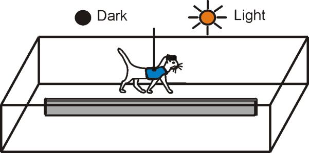
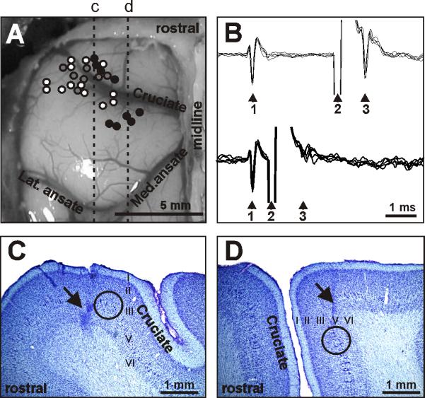
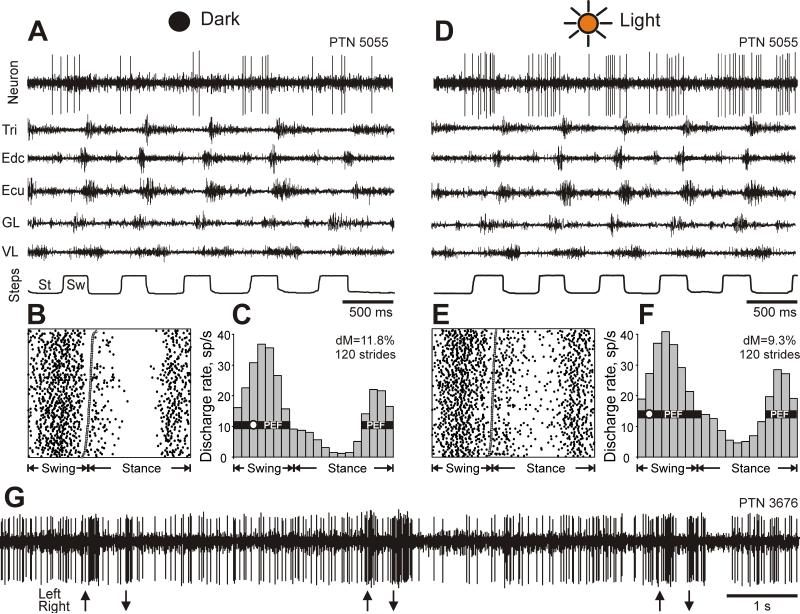
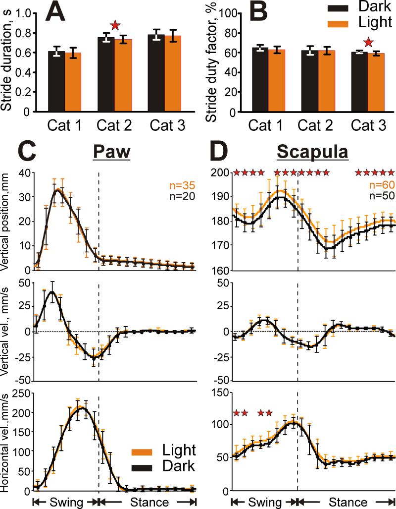
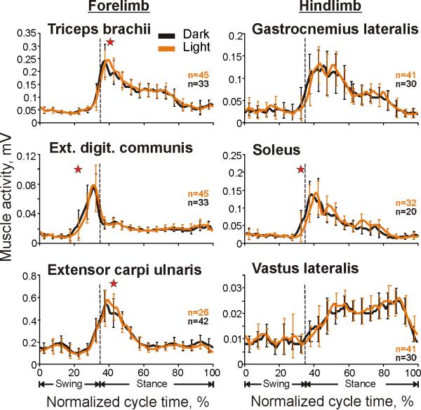
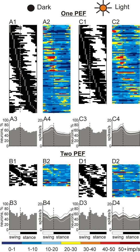
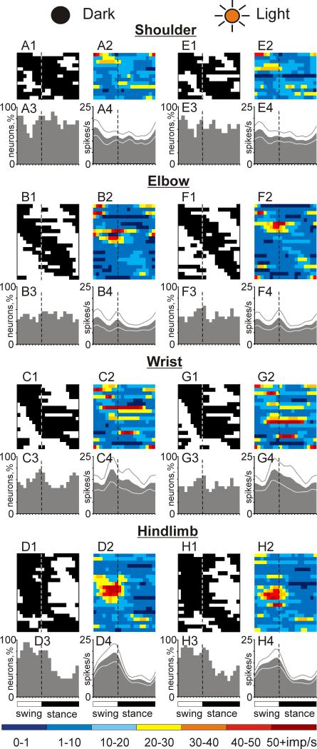
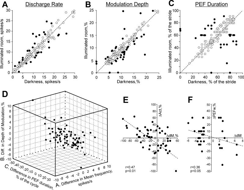
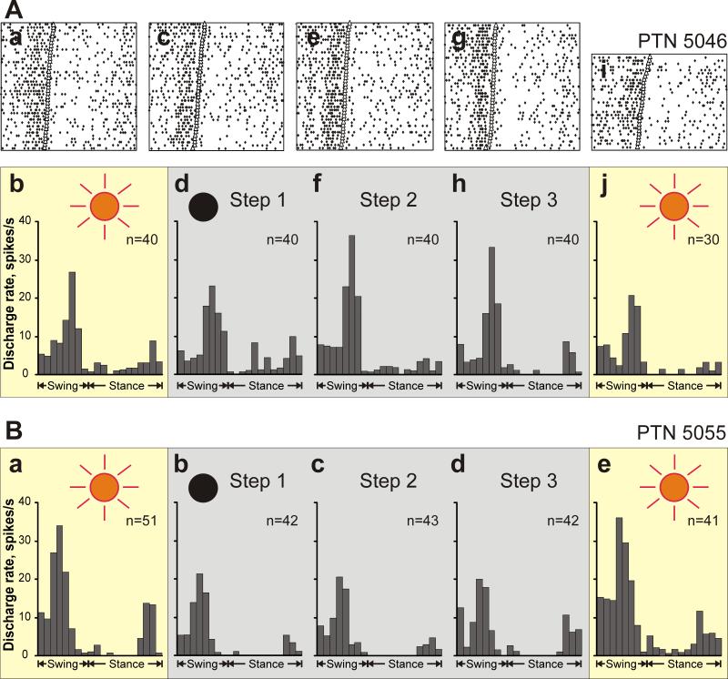
The motor cortex plays a critical role in accurate visually guided movements such as reaching and target stepping. However, the manner in which vision influences the movement-related activity of neurons in the motor cortex is not well understood. In this study we have investigated how the locomotion-related activity of neurons in the motor cortex is modified when subjects switch between walking in the darkness and in light. Three adult cats were trained to walk through corridors of an experimental chamber for a food reward. On randomly selected trials, lights were extinguished for approximately 4s when the cat was in a straight portion of the chamber's corridor. Discharges of 146 neurons from layer V of the motor cortex, including 51 pyramidal tract cells (PTNs), were recorded and compared between light and dark conditions. It was found that while cats' movements during locomotion in light and darkness were similar (as judged from the analysis of three-dimensional limb kinematics and the activity of limb muscles), the firing behavior of 49% (71/146) of neurons was different between the two walking conditions. This included differences in the mean discharge rate (19%, 28/146 of neurons), depth of stride-related frequency modulation (24%, 32/131), duration of the period of elevated firing ([PEF], 19%, 25/131), and number of PEFs among stride-related neurons (26%, 34/131). 20% of responding neurons exhibited more than one type of change. We conclude that visual input plays a very significant role in determining neuronal activity in the motor cortex during locomotion by altering one, or occasionally multiple, parameters of locomotion-related discharges of its neurons.


Figures

Figure 1
Figure 1
Locomotion tasks. The cat walked continuously in a rectangular chamber with two paths. On randomly chosen rounds, lights were turned off upon the cat entering a straight portion of the walkway; they were kept off for approximately four seconds, and were turned back on shortly before the cat reached the turn.
Figure 2
Figure 2
Location and identification of neurons. A: Areas of recording in the left motor cortex. Microelectrode entry points into the cortex were combined from all cats and are shown as circles on the photograph of cat 2 cortex: cat 1, 2, and 3 entry points are depicted by grey, white, and black circles, respectively. Positions of parasagittal sections, whose photomicrographs are shown in C and D are indicated by dotted lines c and d, respectively. B: A collision test determines whether a neuron's response to pyramidal tract stimulation is antidromic. Top: the neuron spontaneously discharges (arrowhead 1) and the pyramidal tract is stimulated approximately 3 ms later (arrowhead 2). The neuron responds with a latency of approximately 1 ms (arrowhead 3). Bottom: the neuron spontaneously discharges (arrowhead 1) and the pyramidal tract is stimulated about 0.5 ms later (arrowhead 2). The neuron does not respond (arrowhead 3) because at 0.5 ms the spontaneous spike was still en route to pyramidal tract, therefore causing the collision, or nullification, of the response. The test confirms that the neurons's response was antidromic, and therefore the neuron proves to be a pyramidal tract projecting neuron (PTN). C: Photomicrograph of a parasagittal section through the lateral pre-cruciate motor cortex (section c). An arrow points to a reference electrolytic lesion in the forelimb representation of the motor cortex. D: Photomicrograph of a parasagittal section through the medial motor cortex (section d). An arrow points to a reference electrolytic lesion in the hindlimb representation of the motor cortex. In C and D: Layers of the cortex are numbered. Groups of giant cells in layer V, which are characteristic for area 4γ and are visible throughout both the pre- and post-cruciate cortex, are encircled. Cresyl violet stain.
Figure 3
Figure 3
A typical example of the activity of a neuron (PTN 5055) and selected right fore- and hindlimb muscles during locomotion in the darkness and light. A: Activity of the neuron and muscles during locomotion in the darkness. Tri, m. triceps brahii (elbow extensor); Edc, m. extensor digitorum communis (wrist and phalanges dorsal flexor); ECU, m. extensor carpi ulnaris (wrist dorsal flexor); GL, m. gastrocnemius lateralis (ankle extensor), VL, m. vastus lateralis (knee extensor). The bottom trace shows the stance (St) and swing (Sw) phases of the step cycle of the right forelimb that is contralateral to the recording site in the cortex. B, C: The activity of the same neuron during locomotion in the darkness is presented as a raster of 120 step cycles (B) and as histograms (C). The duration of step cycles is normalized to 100%. In the raster, the end of swing and the beginning of the stance in each cycle is indicated by an open triangle. In the histogram, the horizontal black bar shows the period of elevated firing (PEF) and the circle indicates the preferred phase as defined in the “Methods” section. The value of dM is stated. D-F: Activities of the same neuron and muscles during locomotion in illuminated room. G: Responses of a neuron to movements of an object in front of the cat (PTN 3676, located in the rostral cruciate sulcus). Arrows pointed up indicate movements from cat's right to left, arrows pointed down indicate movements from cat's left to right.
Figure 4
Figure 4
Kinematics of locomotion in the darkness and light. A: The average duration of the stride of each cat in the two light conditions. B: The average stride duty factor (the percent of the total cycle in which the right forelimb is in the stance phase). C,D: Vertical position (top panel), and vertical (middle panel) and horizontal (bottom panel) velocity of the right paw (C) and scapula (D). Error bars are SDs. Stars denote significant differences in parameters between the conditions (Student's unpaired t test, p<0.05).
Figure 5
Figure 5
Typical examples of EMG activity of selected limb muscles during locomotion in the darkness and light. Each panel shows a representative activity of a muscle, which was averaged over 20-45 strides of each locomotion task, all recorded during one session (see Methods for stride selection). Error bars are SDs. Stars denote significant differences between the conditions (Student's unpaired t test, p<0.05).
Figure 6
Figure 6
Population characteristics of one- and two-PEF neurons during locomotion in the darkness and light. A1, C1: Phase distribution of PEFs of all one-PEF neurons during locomotion in the dark (A1) and under normal illumination (C1). Each row represents the PEF of one cell. A circular mark on each PEF denotes the cell's preferred phase. Neurons are rank-ordered so that those with a preferred phase earlier in the cycle are plotted on the top of the graph. Vertical interrupted lines indicate the end of swing and beginning of stance phase. A2, C2: Corresponding phase distributions of discharge frequencies. The average discharge frequency in each 1/20th portion of the cycle is color-coded according to the scale shown at the bottom of the figure. A3, C3: Proportion of active one-PEF neurons (neurons in their PEF) in different phases of the step cycle during walking in the darkness (A3) and light (C3). A4, C4: The mean discharge rate of one-PEF neurons during walking in the darkness (A4) and under normal illumination (C4). Thin lines show SEM. B1-4 and D1-4 show characteristics of two-PEF neurons locomotion in the darkness (B1-4) and light (D1-4). Designations are similar to those in A,C.
Figure 7
Figure 7
Population characteristics of neurons with different somatosensory receptive fields during locomotion in the darkness and light. A, E: Activity of neurons responsive to movements in the shoulder joint, and/or palpation of back or neck muscles during locomotion in the darkness (A) and under normal illumination (E). A1, E1: Phase distribution of PEFs. A2, E2: Corresponding phase distribution of discharge frequencies. The average discharge frequency in each 1/20th portion of the cycle is color-coded according to the scale shown at the bottom of the figure. A3, E3: Proportion of active neurons (neurons in their PEFs) in different phases of the step cycle. A4, E4: The mean discharge rate. Thin lines show SEM. Vertical interrupted lines denote end of swing and beginning of stance phase. B, F: Activity of neurons responsive to passive movement of the elbow joint or palpation of arm muscles. C, G: Activity of neurons responsive to passive movement in the wrist joint or palpation of muscles on the forearm or paw. D, H: Activity of neurons responsive to stimulation of the hindlimb.
Figure 8
Figure 8
Comparison of activity characteristics of individual neurons between locomotion in the darkness and light. A: Mean discharge frequency averaged over the stride. B: Depth of frequency modulation, dM. C: Duration of PEF, for two-PEF neurons, the combined duration of two PEFs is given. A-C: The abscissa and ordinate of each point show the values of a characteristic of a neuron during locomotion in the darkness and light, respectively. Neurons whose characteristics were statistically significantly different during the two tasks (see Methods) are shown as filled circles; others are shown as open circles. D: Neurons with a statistically significant difference in at least one of the above parameters of the activity between two conditions. The abscissa, ordinate, and applicate of each point show the difference in a discharge characteristic of a neuron between dark and light conditions. The difference is positive if the value of the parameter was larger during locomotion in the light. The star in the middle of the cube denotes the zero point. E: Negative correlation between the relative change in the depth of modulation, dM, and activity, Act. F: Negative correlation between the change in the depth of modulation and duration (width) of PEF, PEFw. In E and F: The abscissa and ordinate of each point show the difference in a discharge characteristic of a neuron between two illumination conditions. The difference is positive if the value of the parameter was larger during locomotion in the light. Only neurons with statistically significant difference in the dM between two walking conditions are shown. The coefficient of correlation (r) is indicated.
Figure 9
Figure 9
Differences in discharge patterns of neurons during locomotion in the darkness and light. A: A schematic presentation of the most frequently observed types of difference in the discharge pattern. In the light, a subtle trough within the one PEF seen during walking in the darkness deepens and divides the PEF into two. The black line shows activity during walking in the darkness, and orange line shows the activity during walking in the light. The vertical dotted line shows end of swing and beginning of stance phase. B: An example activity of a neuron exhibiting this behavior. The dark gray area histogram shows the activity of the neuron during locomotion in the darkness. The orange bar histogram shows the activity during locomotion in the light. To promote visualization of the difference in activities between two tasks, the stride cycle is shown twice. C, D: Same as A, B but showing the second most frequent type of discharge pattern difference. The transition from one- to two-PRF discharge pattern occurred because in the light a new PEF appeared within the former trough. E, F: Same as above but showing the third most frequent type of discharge pattern change in the light from one to two PEFs per cycle: by an increase in activity within a part of the PEF. G, H: same as above but showing the discharge pattern change from two- to one-PEFs per cycle.
Figure 10
Figure 10
Changes in the activity and modulation over the period of darkness. A: An example of a neuron (PTN 5046) that changed the discharge rate and modulation depth during the period of the darkness. Aa,b: The activity of the neuron during locomotion in the light is presented as a raster of 40 step cycles (a) and as a histogram (b). The duration of step cycles is normalized to 100%. In the raster, the end of swing and the beginning of the stance in each cycle is indicated by an open triangle. Ac,d: The activity of the same neuron during the first step made after lights were turned off (Step 1) is shown as a raster of 40 step cycles (c) and as a histogram (d). Ae,f: The activity of the same neuron during the second step after lights were turned off (Step 2) is shown as a raster of 40 step cycles (e) and as a histogram (f). Ag,h: The activity of the neuron during the third step in the darkness (Step 3) is presented as a raster of 40 step cycles (g) and as a histogram (h). Ai,j: The activity of the neuron during the first step made after lights were turned back on is shown as a raster of 30 step cycles (i) and as a histogram (j). B: Histograms of the activity of another neuron (PTN 5055) that had different activities between light and dark conditions but did not change the activity during the period of the darkness.

References

    1. Georgopoulos AP, Kalaska JF, Caminiti R, Massey JT. On the relations between the direction of two-dimensional arm movements and cell discharge in primate motor cortex. J Neurosci. 1982;2:1527–1537. - PMC - PubMed
    1. Kalaska JF. From intention to action: motor cortex and the control of reaching movements. Adv Exp Med Biol. 2009;629:139–178. - PubMed
    1. Scott SH. Inconvenient truths about neural processing in primary motor cortex. J Physiol. 2008;586(5):1217–1224. - PMC - PubMed
    1. Armstrong DM, Drew T. Discharges of pyramidal tract and other motor cortical neurons during locomotion in the cat. J Physiol. 1984a;346:471–495. - PMC - PubMed
    1. Beloozerova IN, Sirota MG. Activity of neurons of the motosensory cortex during natural locomotion in the cat. Neurophysiologia. 1985;17:406–408. - PubMed

Publication types