

Numerical investigation of the effect of cannula placement on thrombosis
- PMID: 23680359
- PMCID: PMC3673905
- DOI: 10.1186/1742-4682-10-35
Numerical investigation of the effect of cannula placement on thrombosis
Abstract
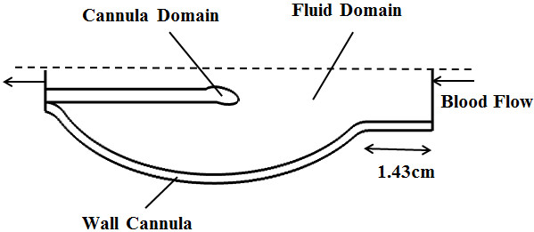
Despite the rapid advancement of left ventricular assist devices (LVADs), adverse events leading to deaths have been frequently reported in patients implanted with LVADs, including bleeding, infection, thromboembolism, neurological dysfunction and hemolysis. Cannulation forms an important component with regards to thrombus formation in assisted patients by varying the intraventricular flow distribution in the left ventricle (LV). To investigate the correlation between LVAD cannula placement and potential for thrombus formation, detailed analysis of the intraventricular flow field was carried out in the present study using a two way fluid structure interaction (FSI), axisymmetric model of a passive LV incorporating an inflow cannula. Three different cannula placements were simulated, with device insertion near the LV apex, penetrating one-fourth and mid-way into the LV long axis. The risk of thrombus formation is assessed by analyzing the intraventricular vorticity distribution and its associated vortex intensity, amount of stagnation flow in the ventricle as well as the level of wall shear stress. Our results show that the one-fourth placement of the cannula into the LV achieves the best performance in reducing the risk of thrombus formation. Compared to cannula placement near the apex, higher vortex intensity is achieved at the one-fourth placement, thus increasing wash out of platelets at the ventricular wall. One-fourth LV penetration produced negligible stagnation flow region near the apical wall region, helping to reduce platelet deposition on the surface of the cannula and the ventricular wall.
Figures


References
-
- WHO Fact sheet No.317.2013. [ http://www.who.int/mediacentre/factsheets/fs317/en/index.html]
-
- Thoennissen NH, Schneider M, Allroggen A, Ritter M, Dittrich R, Schmid C, Scheld HH, Ringelstein EB, Nabavi DG. High level of cerebral microembolization in patients supported with the DeBakey left ventricular assist device. J Thorac Cardiovasc Surg. 2005;130(4):1159–1166. doi: 10.1016/j.jtcvs.2005.02.068. - DOI - PubMed
-
- Schmid C, Jurmann M, Birnbaum D, Colombo T, Falk V, Feltrin G, Garatti A, Genoni M, Gerosa G, Göttel P. Influence of inflow cannula length in axial-flow pumps on neurologic adverse event rate: results from a multi-center analysis. The Journal of Heart and Lung Transplantation: the official publication of the International Society for Heart Transplantation. 2008;27(3):253–260. doi: 10.1016/j.healun.2007.12.007. - DOI - PubMed
Publication types
MeSH terms
LinkOut - more resources
Full Text Sources
Other Literature Sources
Medical
Miscellaneous
