Review
. 2013 May 16;14(5):10242-97.
doi: 10.3390/ijms140510242.

Plant defense against insect herbivores

Affiliations
Review

Plant defense against insect herbivores

Joel Fürstenberg-Hägg et al. Int J Mol Sci. .

Abstract

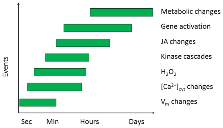
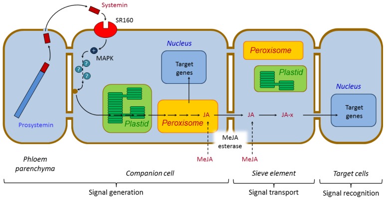
Plants have been interacting with insects for several hundred million years, leading to complex defense approaches against various insect feeding strategies. Some defenses are constitutive while others are induced, although the insecticidal defense compound or protein classes are often similar. Insect herbivory induce several internal signals from the wounded tissues, including calcium ion fluxes, phosphorylation cascades and systemic- and jasmonate signaling. These are perceived in undamaged tissues, which thereafter reinforce their defense by producing different, mostly low molecular weight, defense compounds. These bioactive specialized plant defense compounds may repel or intoxicate insects, while defense proteins often interfere with their digestion. Volatiles are released upon herbivory to repel herbivores, attract predators or for communication between leaves or plants, and to induce defense responses. Plants also apply morphological features like waxes, trichomes and latices to make the feeding more difficult for the insects. Extrafloral nectar, food bodies and nesting or refuge sites are produced to accommodate and feed the predators of the herbivores. Meanwhile, herbivorous insects have adapted to resist plant defenses, and in some cases even sequester the compounds and reuse them in their own defense. Both plant defense and insect adaptation involve metabolic costs, so most plant-insect interactions reach a stand-off, where both host and herbivore survive although their development is suboptimal.


Figures

Figure 1
Figure 1
Evolutionary development of plant classes and insect feeding strategies. The evolutionary developments of new classes of plants are shown accompanied by new feeding strategies due to the interactions between plants and insects. The numerals on top refer to the numbers of feeding strategies present. Below are the dominating feeding strategies, followed by the first occurrences of certain feeding modes. The geographic time scale and colors are obtained from the International Commission of Stratigraphy, where Sil—Silurian, Devon—Devonian, Carbon—Carboniferous, Perm—Permian, Tria—Triassic, Jura—Jurassic, Creta—Cretaceous, Pale—Paleogene, Neo—Neogene and Qua—Quarternary (Adapted from [–4]).
Figure 2
Figure 2
Structures of oral insect secretions. (a) Volicitin, N-(17-hydroxylinolenoyl)-l-Gln; (b) Inceptin, proteolytic peptides of the chloroplastic ATP synthase γ-subunit; (c) Caeliferin A16:1, (E)-2,16 disulfooxy-6-hexadecenoic acid; and (d) Bruchin c, (Z)-9-tetracosene-1,24-diol bis-(3-hydroxypropanoate)ester.
Figure 3
Figure 3
Events in plants after feeding by insect herbivores. Changes in the transmembrane potential (Vm) appear immediately upon herbivory damage and are tightly followed by changes in the intracellular Ca2+ concentration and generation of H2O2. Kinases and the phytohormone jasmonic acid (JA) are detectable within minutes. After roughly 1 h, gene activation followed by metabolic changes is seen (Adapted from [44]).
Figure 4
Figure 4
Model of systemic signaling and activation of defense genes in response to wounding by insect attack. After wounding, the systemin peptide is released from the C-terminal end of its precursor prosystemin by proteolytic processing. Systemin then enters the apoplast, where it binds to a membrane-bound receptor (SR160) to initiate an intracellular signaling cascade. The cascade includes the activities of a MAP kinase (MAPK), and a couple of unknown intermediates, leading to the release of polyunsaturated fatty acids (PUFAs) by phospholipases, from the membranes. The biosynthesis of JA takes place in the chloroplast and peroxisome within the companion cell, after which it might be transported long distances via the phloem. Plasmodesmatal connections between different cell types are shown as brown pipes. JA or a covalently modified form of JA (JA-x; such as JA-Ile) activates target gene expression in distal undamaged leaves. Esterases may convert exogenous MeJA to JA upon diffusion of MeJA across membranes. For simplicity, cell types presumed to be involved in phloem unloading of the signal are not shown. Mobile signals are shown in red and nonmobile signals in blue. Putative steps are denoted with dashed arrows (Adapted from [83,88]).
Figure 5
Figure 5
Biosynthesis of jasmonic acid in the chloroplast and peroxisome. Polyunsaturated fatty acids (18:3 and 16:3) released from the cell, chloroplast and/or thylakoid membrane are precursors for the biosynthesis of jasmonic acid (JA). Within the chloroplast, cis-(+)-12-oxo-phytodienoic acid (cisOPDA) and dinor-OPDA (dnOPDA) are formed via the octa- and hexadecanoid pathways. After transport into the peroxisome, OPDA (dnOPDA) is reduced to OPC8:0 (OPC6:0) and undergoes three (two) cycles of β-oxidation that results in the production of (+)-7-epi-jasmonic acid. The reactions are catalyzed by lipoxygenases (LOX), allene oxide synthase (AOS), allene oxide cyclase (AOC), ATP-binding cassette (ABC) transporter COMATOSE (CTS/PXA1/PED3), 12-oxophyto-dienoate reductase (OPR3), OPC CoA ligase1 (OPCL), acyl-thioesterase (ACH), 3-ketoacyl-CoA thiolase [132], acyl-CoA oxidase (ACX) and a multifunctional protein (MFP). Enzymes are shown in blue. Arrows show the well characterized reactions, whereas dashed arrows show steps that are still hypothetical, and for which the corresponding enzymes remain to be identified. (Adapted from [117,133]).
Figure 6
Figure 6
Model of jasmonate regulation of defense responsive genes. (a) In the resting state, in the absence of JA, JAZ proteins will bind to transcription factors, such as MYC2, and prevent expression of the JA-responsive genes; (b) In the active state, wounding promotes JA biosynthesis, resulting in accumulation of (+)-7-epi-jasmonyl-l-isoleucine ((+)-7-epi-JA-l-Ile). The hormone will bind to and stabilize the COI1 F-box subunit of the COI1 E3 ubiquitin ligase enzymatic complex (SCFCOI1), which in its turn bind to the Jas motif (J) of the Jasmonate ZIM-domain protein (JAZ), leading to ubiquitination and subsequent degradation by the 26S proteasome (26S). The transcription factors will now be free to recruit the RNA polymerase II transcriptional machinery to the promoter of the JA-responsive genes, assisted by universal adaptors, such as the Mediator complex; (c) In the presence of (+)-7-epi-JA-l-Ile and the absence of gibberellic acids (GAs), stabilized DELLA proteins (DELLA) will compete with MYC2 for binding of JAZ, thereby releasing MYC2 for activation of the JA-response; (d) If GAs are present, they will bind to DELLA and trigger degradation. This will liberate JAZ, promote the formation of the JAZ-MYC complex, and thereby repress the expression of JA-responsive gene (Adapted from [131,151]).
Figure 7
Figure 7
Structures of charged oligosaccharides inducing plant defense-responses. (a) The elicitor oligo (α-1,4) galacturonic acid [97] is formed by the action of polygalacturonase (PG) on plant cell wall pectins; and (b) chitosan, an oligomer of β-1,4-linked glucosamine, can also act as an elicitor (Adapted from [83]).
Figure 8
Figure 8
Structures of plant bioactive natural products. (a) nicotine, a true alkaloid derived from aspartate and ornithine; (b) DIMBOA, a benzoxazinoide derived from indole-3-glycerol phosphate; (c) Dhurrin, a cyanogenic glucoside derived from tyrosine, (d) Sinalbin, a glucosinolate derived from tyrosine; (e) Canavanine, a nonprotein amino acid derived from l-homoserine; (f) Salicylic acid, a benzoic acid derived phenol; and (g) S(−)-limonene, a terpenoid derived from geranyl pyrophosphate [189].

Similar articles

Cited by

References

    1. Ehrlich P.R., Raven P.H. Butterflies and plants: A study in coevolution. Evolution. 1964;18:586–608.
    1. Labandeira C.C. Insect mouthparts: Ascertaining the paleobiology of insect feeding strategies. Annu. Rev. Ecol. Syst. 1997;28:153–193.
    1. Niklas K.J., Tiffney B.H., Knoll A.H. Patterns in Vascular Land Plant Diversification: An Analysis at the Species Level. In: Valentine J.W., editor. Phanerozoic Diversity Patterns: Profiles in Macroevolution. Princeton University Press; Princeton, NJ, USA: 1985. pp. 97–128.
    1. Schönenberger J., Friis E.M., Matthews M.L., Endress P.K. Cunoniaceae in the cretaceous of europe: Evidence from fossil flowers. Ann. Bot. 2001;88:423–437.
    1. Schaller A., Howe G.A. Direct Defenses in Plants and Their Induction by Wounding and Insect Herbivores. In: Schaller A., editor. Induced Plant Resistance to Herbivory. Springer Science+Business Media; Berlin, Germany: 2008. pp. 7–29.

Publication types

Substances