. 2013 May 16;4(5):e635.
doi: 10.1038/cddis.2013.159.

SIRT1 activation enhances HDAC inhibition-mediated upregulation of GADD45G by repressing the binding of NF-κB/STAT3 complex to its promoter in malignant lymphoid cells

Affiliations

SIRT1 activation enhances HDAC inhibition-mediated upregulation of GADD45G by repressing the binding of NF-κB/STAT3 complex to its promoter in malignant lymphoid cells

A Scuto et al. Cell Death Dis. .

Abstract

We explored the activity of SIRT1 activators (SRT501 and SRT2183) alone and in combination with panobinostat in a panel of malignant lymphoid cell lines in terms of biological and gene expression responses. SRT501 and SRT2183 induced growth arrest and apoptosis, concomitant with deacetylation of STAT3 and NF-κB, and reduction of c-Myc protein levels. PCR arrays revealed that SRT2183 leads to increased mRNA levels of pro-apoptosis and DNA-damage-response genes, accompanied by accumulation of phospho-H2A.X levels. Next, ChIP assays revealed that SRT2183 reduces the DNA-binding activity of both NF-κB and STAT3 to the promoter of GADD45G, which is one of the most upregulated genes following SRT2183 treatment. Combination of SRT2183 with panobinostat enhanced the anti-growth and anti-survival effects mediated by either compound alone. Quantitative-PCR confirmed that the panobinostat in combination with SRT2183, SRT501 or resveratrol leads to greater upregulation of GADD45G than any of the single agents. Panobinostat plus SRT2183 in combination showed greater inhibition of c-Myc protein levels and phosphorylation of H2A.X, and increased acetylation of p53. Furthermore, EMSA revealed that NF-κB binds directly to the GADD45G promoter, while STAT3 binds indirectly in complexes with NF-κB. In addition, the binding of NF-κB/STAT3 complexes to the GADD45G promoter is inhibited following panobinostat, SRT501 or resveratrol treatment. Moreover, the combination of panobinostat with SRT2183, SRT501 or resveratrol induces a greater binding repression than either agent alone. These data suggest that STAT3 is a corepressor with NF-κB of the GADD45G gene and provides in vitro proof-of-concept for the combination of HDACi with SIRT1 activators as a potential new therapeutic strategy in lymphoid malignancies.


Figures

Figure 1
Figure 1
SIRT1 activators inhibit viability of human malignant lymphoid B-cell lines. (a) Reh cells and (b) Nalm-6 cells were treated with the indicated concentrations of SRT2183 or SRT501 for 24, 48, or 72 h. Following this, the percentage of cell viability was determined by MTS assay. Values represented as graphs are the mean of three independent experiments with S.D. (c) Cells were treated with the indicated concentrations of SRT2183 for 48 h. Following this, the percentage of cell viability was determined by MTS assay. (d) Peripheral blood mononuclear cells from a healthy donor were treated in culture with the indicated concentrations of SRT2183 or SRT501 for 48 h. The percentage of viable cells was determined by DIMSCAN analysis. Values represent the percentages of cell viability normalized to that of the untreated cells and are the mean of three separate treatments
Figure 2
Figure 2
SIRT1 activators induce apoptosis of human Ph ALL cell lines. (a) Reh cells and (b) MOLT-4 cells were treated with the indicated concentrations of SRT2183 or SRT501 for 48 h. Following this, the percentage of apoptotic cells was determined by flow cytometry using Annexin V/propidium iodide staining. Values represented as bar graphs are the mean of three independent experiments with S.D.
Figure 3
Figure 3
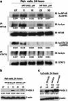
SIRT1 activators lead to deacetylation of both STAT3 and NF-κB p65 and induce phosphorylation of H2A.X. Cells were treated with the indicated concentrations of SRT2183 or SRT501 for 24 h. Following this, (a) immunoprecipitation (IP) and subsequent immunoblotting (IB) were performed on the lysates from Reh cells to analyze acetyl-STAT3 and acetyl-NF-κB levels. The levels of total STAT3 and total NF-κB served as loading controls. Results are representative of three independent experiments. Western blot analysis of phospho-H2A.X protein was performed on the lysates from (b) Reh or (c) Ly3 cells 24 h post treatment. The levels of β-actin protein served as loading controls. Results are representative of three independent experiments
Figure 4
Figure 4
Combination of SRT2183 with panobinostat induces more apoptosis than either compound alone in Reh cells. (a) Reh cells were treated for 48 h with the indicated concentrations of SRT2183, panobinostat, or a combination of these drugs. Following this, the percentage of cell viability was determined by MTS assay. Values represented as graphs are the mean of three independent experiments with S.D. (b) Reh cells were treated for 48 h with the indicated concentrations of SRT2183, panobinostat, or a combination of these drugs. Following this, the percentage of late apoptotic cells or total (early plus late) apoptosis was determined by flow cytometry using Annexin V/propidium iodide staining. Values represented as bar graphs are the mean of three independent experiments with the S.D. (c) Ly3 cells were treated for 48 h with the indicated concentrations of SRT2183, panobinostat, or a combination of these drugs. Following this, the percentage of cell viability was determined by MTS assay. Values represented as graphs are the mean of three independent experiments with S.D. (d) Peripheral blood mononuclear cells from a healthy donor were treated in culture for 48 h with the indicated concentrations of SRT2183, panobinostat, or a combination of these drugs. The percentage of viable cells was determined by DIMSCAN analysis. Values represent the percentages of cell viability normalized to that of the untreated cells and are the mean of three separate treatments. Statistical significance is relative to the single agent treatment inducing higher effect. NS, not significant, *P<0.05, **P<0.01, ***P<0.001
Figure 5
Figure 5
Combination of SRT2183 with panobinostat induces greater phosphorylation of H2A.X and acetylation of H4 in ALL cell lines. (a) Reh cells and (b) NALM-6 cells were treated for 24 h with the indicated concentrations of SRT2183, panobinostat, or a combination of these drugs. Following this, western blot analysis of phospho-H2A.X and acetyl-H4 proteins was performed on the lysates from those cells. The levels of β-actin protein served as loading controls. Results are representative of three independent experiments
Figure 6
Figure 6
Combination of SRT2183 with panobinostat induces greater upregulation of GADD45G and GADD45A genes than either compound alone in pre-B ALL cell lines. (a) Reh cells, (b) NALM-6 cells and (c) Ly3 cells were treated for 24 h with the indicated concentrations of panobinostat and SIRT1 activators as single agents or in combination with panobinostat. Following this, TaqMan real-time PCR was performed. The mRNA levels were normalized to levels of β-actin mRNA. Values represented as bar graphs are the mean of three independent experiments with S.D. Data are expressed as 2^ddCt. (d) Reh cells were transfected with Cy3-labeled GADD45G siRNA or negative control siRNA and treated, 48 h later, with the indicated concentrations of SRT2183, panobinostat ,or a combination of these drugs. At 48 h after treatment, the percentage of apoptotic cells was determined in the gated Cy3-positive cell population, by flow cytometry using Annexin V/DAPI staining. Values represented as bar graphs are the mean of three independent experiments with S.D.
Figure 7
Figure 7
Effects of SIRT1 activators alone or in combination with panobinostat on the binding of NF-κB and STAT3 to the GADD45G promoter. (a) Following treatment with 10 μM SRT2183 for 24 h, ChIP assay was performed on Reh and Ly3 cells. DNAs purified from the sheared cross-linked chromatin immunoprecipitated with anti-acetyl-STAT3 or anti-acetyl-NF-κB antibodies were used as TaqMan real-time PCR templates to amplify the GADD45G promoter region from −567 to +6. Inputs were used to normalize. Values represented as bar graphs are the mean of three independent experiments with S.D. (b) Competition EMSA: Effect of unlabeled (‘cold') oligonucleotides G1, G2, and G3 on NF-κB DNA-binding activity. Lanes 1–7: Nuclear extract from lipopolysaccharide LPS-stimulated macrophages. Lanes 8–14: Nuclear extract from Ly3 cells. ‘Cold' oligonucleotides G1–G3 and C1 were added in 100-fold molar excess, C2 was added 1 : 1, as compared with control 32P-labeled NF-κB oligo C2. Lanes 2 and 9 contain NF-κB antibodies (supershift). (c) DNA-binding activity (EMSA assay) of NF-κB to 32P-labeled NF-κB consensus sequences G1–G3 (lanes 1–9). Lanes 10–15: Control NF-κB oligonucleotides. Lanes 1–15: Nuclear extract from Ly3 cells. NF-κB and STAT3 antibodies were used in supershifts (lanes 2, 5, 8, 11, 14 and lanes 3, 6, 9, 12, 15, respectively). (d) Effect of compound-treated Ly3 cells on NF-κB DNA-binding activity (EMSA assay). Ly3 cells were treated with HDACi, SIRTa or HDACi+SIRTa as indicated. Nuclear extract of untreated (lanes 1–3) and treated cells (lanes 4–10) was isolated 24 h post treatment and incubated with 32P-labeled NF-κB oligonucleotide G1 before EMSA analysis. NF-κB and STAT3 antibodies were used in supershift (lanes 2 and 3, respectively)
Figure 8
Figure 8
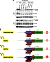
A model to explain the greater upregulation of GADD45G mediated by the combination of a SIRT1 activator with a class I, II, and IV HDAC inhibitor. (a) Ly3 cells were treated with the indicated concentrations of panobinostat, and SIRT1 activators as single agents or in combination with panobinostat for 24 h. Following this, immunoprecipitation (IP) and subsequent immunoblotting (IB) were performed on the cell lysates to analyze acetyl-STAT3 and acetyl-NF-κB levels. The levels of total STAT3 and total NF-κB served as loading controls. (b) Predicted acetylation status of histones and STAT3/NF-κB complexes at the GADD45G promoter in untreated cells (i) and in cells treated with an HDACi (ii), a SIRT1a (iii), and the combination of an HDACi with a SIRT1a (iv)

Similar articles

Cited by

References

    1. Stimson L, Wood V, Khan O, Fotheringham S, La Thangue NB. HDAC inhibitor-based therapies and haematological malignancy. Ann Oncol. 2009;20:1293–1302. - PubMed
    1. Piekarz RL, Frye R, Turner M, Wright JJ, Allen SL, Kirschbaum MH, et al. Phase II multi-institutional trial of the histone deacetylase inhibitor romidepsin as monotherapy for patients with cutaneous T-cell lymphoma. J Clin Oncol. 2009;27:5410–5417. - PMC - PubMed
    1. Kirschbaum M, Frankel P, Popplewell L, Zain J, Delioukina M, Pullarkat V, et al. Phase II study of vorinostat for treatment of relapsed or refractory indolent non-Hodgkin's lymphoma and mantle cell lymphoma. J Clin Oncol. 2011;29:1198–1203. - PMC - PubMed
    1. Frye RA. Characterization of five human cDNAs with homology to the yeast SIR2 gene: Sir2-like proteins (sirtuins) metabolize NAD and may have protein ADP-ribosyltransferase activity. Biochem Biophys Res Commun. 1999;260:273–279. - PubMed
    1. Finkel T, Deng CX, Mostoslavsky R. Recent progress in the biology and physiology of sirtuins. Nature. 2009;460:587–591. - PMC - PubMed

Publication types

MeSH terms