Review
. 2013 Jun;14(6):509-19.
doi: 10.1038/embor.2013.49. Epub 2013 May 17.

Mechanosensitivity and compositional dynamics of cell-matrix adhesions

Affiliations
Review

Mechanosensitivity and compositional dynamics of cell-matrix adhesions

Herbert B Schiller et al. EMBO Rep. 2013 Jun.

Abstract

Cells perceive information about the biochemical and biophysical properties of their tissue microenvironment through integrin-mediated cell-matrix adhesions, which connect the cytoskeleton with the extracellular matrix and thereby allow cohesion and long-range mechanical connections within tissues. The formation of cell-matrix adhesions and integrin signalling involves the dynamic recruitment and assembly of an inventory of proteins, collectively termed the 'adhesome', at the adhesive site. The recruitment of some adhesome proteins, most notably the Lin11-, Isl1- and Mec3-domain-containing proteins, depends on mechanical tension generated by myosin II-mediated contractile forces exerted on cell-matrix adhesions. When exposed to force, mechanosensitive adhesome proteins can change their conformation or expose cryptic-binding sites leading to the recruitment of proteins, rearrangement of the cytoskeleton, reinforcement of the adhesive site and signal transduction. Biophysical methods and proteomics revealed force ranges within the adhesome and cytoskeleton, and also force-dependent changes in adhesome composition. In this review, we provide an overview of the compositional dynamics of cell-matrix adhesions, discuss the most prevalent functional domains in adhesome proteins and review literature and concepts about mechanosensing mechanisms that operate at the adhesion site.


Conflict of interest statement

The authors declare that they have no conflict of interest.

Figures

Figure 1
Figure 1
Reciprocal feedback connections at cell–matrix adhesions involve mechanosensitive modules. Both the chemistry and the mechanical properties of the ECM are controlled by pericellular mechanisms such as matrix crosslinking, fibrillogenesis and proteolysis, affecting integrin use and activation. These properties, and the integrins that become engaged, influence the composition of connectors and signal transducers that are recruited to cell–matrix adhesion sites leading to specific signalling and regulation of cytoskeletal components. Myosin II-mediated pulling on stiff ECM generates tension that is distributed along ECM fibrils, the cell–matrix adhesome and the cytoskeletal filaments, which promotes the partial unfolding or conformational rearrangement of mechanosensitive adhesome proteins and ECM ligands. Such allosteric mechanisms lead to the recruitment of new proteins into tensioned cell–matrix adhesions resulting in the activation of signalling pathways as well as structural reinforcement and stabilization of adhesion sites, which in turn feed back on various signalling events. Furthermore, biophysical mechanisms such as catch-bonding of, for example, integrin–ligand and F-actin–myosin II interactions are involved in cellular mechanosensing. ECM, extracellular matrix; Erk, extracellular signal-regulated kinase; F-actin, filamentous actin; GAP, GTPase-Activating Protein; GEF, guanine nucleotide exchange factor; GPV, platelet glycoprotein V; Larg, leukaemia-associated RhoGEF; RHAMM, receptor for hyaluronan-mediated motility; Src, proto-oncogene c-Src.
Figure 2
Figure 2
Myosin II-mediated adhesion maturation involves the zinc-finger type LIM domain. (A) A mouse fibroblast plated on fibronectin immunostained for the focal adhesion protein paxillin, the lamellipodium protein cortactin and F-actin. Nascent adhesions (arrows) form in the protruding, cortactin-positive lamellipodium and are not linked to F-actin bundles. By contrast, focal adhesions (arrowheads) are stabilized by their linkage to contractile F-actin bundles. (B) In the course of adhesion maturation, myosin II-mediated pulling through F-actin bundles induces the recruitment of specific proteins such as LIM-domain-containing proteins to cell–matrix adhesions. The box in the panel on the right shows proteins that are enriched in nascent adhesions (gene names in blue) or focal adhesions (gene names in red; LIM-domain proteins are marked with asterisks). Data selected from references [8,52]. FA, focal adhesion; F-actin, filamentous actin; LIM, Lin11, Isl1, Mec3; NA, nascent adhesion.
Figure 3
Figure 3
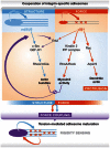
Integrin-specific adhesomes cooperate for rigidity sensing. α5β1-integrins adhere to fibronectin, assemble kindlin 2- and Ilk-rich small peripheral adhesions in a myosin II-independent manner. The protein assembly in α5β1-containing adhesions activates Rac1, Wave and Arp2/Arp3-driven actin polymerization to induce membrane protrusions, and RhoA/Rock-mediated myosin II activation to induce tension. This tension increases the adhesion lifetime of αv-class integrins bound to ligand on stiff substrates, which reinforces and stabilizes focal adhesions. GEF-H1 is recruited by αv-class integrins to focal adhesions, which reinforces RhoA/myosin II in an α5β1-dependent manner, and increase RhoA activity to promote mDia-mediated stress fibre formation. The combination of αv-class integrin-mediated structure (focal adhesion anchoring and stress fibre formation) with the α5β1-mediated force generation (myosin II activity) constitutes a synergistic system, which is important for adapting cellular contractility and architecture to the rigidity of fibronectin-based microenvironments. Scheme adapted from reference [52]. Arp2/3, actin-related protein 2/3; Erk, extracellular signal-regulated kinase; GEF-H1, guanine nucleotide exchange factor H1; IPP, Ilk, Pinch, Parvin.
Figure 4
Figure 4
Proteomic analysis of cell–matrix adhesomes. (A) Workflow for quantitative mass spectrometry and bioinformatic analysis of purified cell–matrix adhesions, as described in [8]. (B) The adhesome components determined by proteomics were assessed for the abundance of distinct domains (PFAM, PROSITE). The pie chart depicts the quantitative distribution of protein domains. CAMSAP, calmodulin-regulated spectrin-associated protein; EGF, epidermal growth factor; FA, focal adhesion; FERM, 4.1 protein, ezrin, radixin, moesin; GAP, GTPase-activating protein; IRS-1, insulin receptor substrate 1; KEGG, Kyoto Encyclopedia of Genes and Genomes; LC, liquid chromatography; LIM, Lin11-, Isl1- and Mec3; MS, mass spectrometry; PAGE, polyacrylamide gel electrophoresis; PDZ, PSD95, Dlg1, zo-1; PFAM, protein family; PH, Pleckstrin homology; PTB, phosphotyrosine binding; SDS, sodium dodecyl sulfate; SH2/3, src homology 2/3; SILAC, stable isotope labeling by/with amino acids in cell culture; WD, tryptophan-aspartic acid 40 repeat; WH1, WASP homology 1.

References

    1. Hoffman BD, Grashoff C, Schwartz MA (2011) Dynamic molecular processes mediate cellular mechanotransduction. Nature 475: 316–323 - PMC - PubMed
    1. Ingber DE (1997) Tensegrity: the architectural basis of cellular mechanotransduction. Annu Rev Physiol 59: 575–599 - PubMed
    1. Oda H, Takeichi M (2011) Evolution: structural and functional diversity of cadherin at the adherens junction. J Cell Biol 193: 1137–1146 - PMC - PubMed
    1. Hynes RO (2002) Integrins: bidirectional, allosteric signaling machines. Cell 110: 673–687 - PubMed
    1. Kanchanawong P, Shtengel G, Pasapera AM, Ramko EB, Davidson MW, Hess HF, Waterman CM (2010) Nanoscale architecture of integrin-based cell adhesions. Nature 468: 580–584 - PMC - PubMed

Publication types

LinkOut - more resources