. 2013 Aug;31(8):1683-95.
doi: 10.1002/stem.1419.

KIT receptor gain-of-function in hematopoiesis enhances stem cell self-renewal and promotes progenitor cell expansion

Affiliations

KIT receptor gain-of-function in hematopoiesis enhances stem cell self-renewal and promotes progenitor cell expansion

Shayu Deshpande et al. Stem Cells. 2013 Aug.

Abstract

The KIT receptor tyrosine kinase has important roles in hematopoiesis. We have recently produced a mouse model for imatinib resistant gastrointestinal stromal tumor (GIST) carrying the Kit(V558Δ) and Kit(T669I) (human KIT(T670I) ) mutations found in imatinib-resistant GIST. The Kit(V558Δ;T669I/+) mice developed microcytic erythrocytosis with an increase in erythroid progenitor numbers, a phenotype previously seen only in mouse models of polycythemia vera with alterations in Epo or Jak2. Significantly, the increased hematocrit observed in Kit(V558Δ;T669I/+) mice normalized upon splenectomy. In accordance with increased erythroid progenitors, myeloerythroid progenitor numbers were also elevated in the Kit(V558Δ;T669I/+) mice. Hematopoietic stem cell (HSC) numbers in the bone marrow (BM) of Kit(V558Δ;T669I/+) mice were unchanged in comparison to wild-type mice. However, increased HSC numbers were observed in fetal livers and the spleen and peripheral blood of adult Kit(V558Δ;T669I/+) mice. Importantly, HSC from Kit(V558Δ;T669I/+) BM had a competitive advantage over wild-type HSC. In response to 5-fluorouracil treatment, elevated numbers of dividing Lin(-) Sca(+) cells were found in the Kit(V558Δ;T669I/+) BM compared to wild type. Our study demonstrates that signaling from the Kit(V558Δ;T669I/+) receptor has important consequences in hematopoiesis enhancing HSC self-renewal and resulting in increased erythropoiesis.

Keywords: Erythroid progenitors; Hematopoietic stem cells; Self-renewal; Signal transduction.


Figures

Figure 1
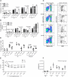
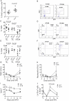
Figure 1. KitV558Δ;T669I/+ mice develop erythrocytosis which is remedied by splenectomy
Total BM and spleen (SP) cells were prepared for staining with Ter119 and CD71 antibodies for flow cytometry (A-B) Graphical representation of flow cytometry results show increased frequency and total Ter119+CD71+ cells, especially R1 erythroid progenitor population in the KitV558Δ;T669I/+ (GTK) BM and spleen. Frequency and total numbers of A and B populations are also significantly increased in the mutant spleen compared to wild-type (WT) (n=4-9). (C) Representative flow cytometric histograms of bone marrow and spleen cells show cells labeled with Ter119 and CD71 antibodies. The frequency of R1 and Ter119hi populations in total BM or spleen cells is depicted. Ter119hi population was further analyzed into A, B and C subsets and numbers indicate frequency of cells within total BM or spleen cells. (D) BM and spleen cells were stained with cell surface markers Ter119 and CD71 and processed for Ki67-DAPI staining. Erythroid R1 progenitor cycling measured by Ki67 flow cytometry in the BM and spleen revealed increased cycling of progenitors in the KitV558Δ;T669I/+ spleen compared to wild-type but no change was observed in the BM of the two groups (n=3-5). (E) In order to assess the contribution of the spleen toward increased erythropoiesis in the KitV558Δ;T669I/+, wild-type and KitV558Δ;T669I/+ mice were splenectomized. Hematocrit (HCT) levels two weeks post-surgery were significantly reduced in KitV558Δ;T669I/+ splenectomized mice compared to pre-surgical values, while wild-type hematocrit levels did not decrease significantly (n=5) (F) Flow cytometric analysis of BM from wild-type and KitV558Δ;T669I/+ by Ter119 and CD71 staining at 8 weeks post-splenectomy showed similar numbers of erythroid R1 progenitors in the BM of KitV558Δ;T669I/+ compared to wild-type splenectomized mice (n=3). Data is representative of at least two independent experiments. n-indicates number of mice per genotype. Values are mean±SEM.
Figure 2
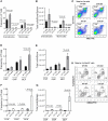
Figure 2. Expansion of the myeloid lineage in the adult BM and spleen of KitV558Δ;T669I/+ mice
Cells from the BM and spleen were stained with Gr1 and Mac1 antibodies and analyzed by flow cytometry. (A) Analysis of Mac1+-Gr1+ population in the BM and spleen showed significantly increased frequency of granulocyte and monocyte populations in KitV558Δ;T669I/+ mice compared to wild-type (n=4-6). (B) Total cell numbers of granulocyte and monocyte cells is significantly increased in the KitV558Δ;T669I/+ spleen compared to wild-type (n=4-6). (C) Representative staining profiles show frequency of granulocytes and monocytes in total BM and spleen. (D,E) BM cells were stained against lineage markers and Lin-Kit+Sca- cells were distinguished into CMP, GMP and MEP subsets according to their CD34 and FcγRII/III surface expression. Flow cytometric analysis showed increased frequency of MEP in the mutant BM compared to wild-type. Total MEP numbers in the mutant BM were increased 5-fold over wild-type (n=3-4). (F,G) Spleen cells from wild-type and mutant stained for myeloid progenitor markers showed increased frequency of CMP, GMP and MEP subsets in the KitV558Δ;T669I/+ spleen. While total CMP numbers were significantly increased in the mutant spleen, MEP numbers were remarkably (20-fold) higher in KitV558Δ;T669I/+ compared to wild-type (n=3-4). (H) Representative flow cytometry plot for myeloerythroid progenitors. Lin-Sca-Kit+ (LSK)cells were divided into GMP, CMP and MEP subsets. Numbers indicate frequency of cells in total BM and spleen. Data is representative of at least two independent experiments. Values are mean±SEM. n-indicates number of mice.
Figure 3
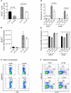
Figure 3. Lymphopoiesis in KitV558Δ;T669I/+ mice
(A-B) BM and spleen cells were stained with B220 antibody and analyzed by flow cytometry. The frequency of B220+ cells was significantly reduced in the KitV558Δ;T669I/+ BM and spleen compared to WT (n=3-5), however total cell numbers were not significantly reduced compared to WT (n=3-5). (C) Representative staining profile shows frequency of B220+ cells in BM and spleen. (D,E) The frequency of CD4+ and CD8+ cells were reduced in the KitV558Δ;T669I/+ BM and spleen, however total cell numbers were not significantly reduced compared to WT (n=3-5). (F) Representative staining profile shows frequency of CD4+ and CD8+ subsets in BM and spleen. Data is representative of at least two independent experiments. Values are mean±SEM. n-indicates number of mice.
Figure 4
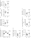
Figure 4. Analysis of the stem cell compartment in wild-type and KitV558Δ;T669I/+ mice
(A-B) Total BM and spleen cells were stained against lineage cocktail and gated as Lin-Kit+Sca+ (LSK). LSK frequency and total cell numbers were not significantly altered in the KitV558Δ;T669I/+ BM compared to wild-type BM but in the KitV558Δ;T669I/+ spleen total LSK numbers were 11-fold elevated compared to wild-type spleen (n=5). (C-D) LT-HSC defined as Lin-Sca+Kit+CD150+CD34- were significantly higher in frequency and total cell numbers were 6-fold higher in KitV558Δ;T669I/+ spleen compared to wild-type spleen. LT-HSC numbers were not significantly different in the BM of the two mice (n=4-5). (E) The frequency of LT-HSC numbers was increased in the peripheral blood of KitV558Δ;T669I/+ compared to wild-type mice (n=4). (F) Representative staining profiles for LSK and LT-HSC from BM and spleen. Lineage- cells were electronically gated into LSK subset. LSK cells were subdivided into LT-HSC, multipotent progenitors (MPP) and lymphoid-primed multipotent progenitors (LMPP). Numbers represent frequency of cells in total BM or spleen. Data is representative of at least three independent experiments. Values are mean±SEM. n-indicates number of mice.
Figure 5
Figure 5. Analysis of fetal livers from wild-type and KitV558Δ;T669I/+ mice
(A) The total number of nucleated cells is higher in KitV558Δ;T669I/+ fetal livers at 14.5 dpc. (B,C) LSK frequency and numbers were not significantly different in KitV558Δ;T669I/+ mice compared to WT. (D,E)LT-HSC frequency and number were significantly higher in KitV558Δ;T669I/+ fetal livers compared to wild-type. (F) Analysis of erythroid progenitors by staining fetal livers against Ter119 and CD71 antibodies revealed significantly higher R1 progenitors in KitV558Δ;T669I/+ compared to wild-type fetal livers, while the erythroid subsets A, B and C did not vary significantly between the two groups. (G,H) Lineage analysis revealed no significant difference in B-lymphoid (B220+), or myeloid (Gr1+) cell numbers between KitV558Δ;T669I/+ and wild-type mice. Graphs A-H: data is representative of at least two independent experiments. n=6-8 fetal livers per group. Values are mean±SEM.
Figure 6
Figure 6. Analysis of HSC self-renewal from wild-type and KitV558Δ;T669I/+ BM and spleen
(A,B) CD45.1 mice were lethally irradiated and transplanted with CD45.1 whole BM cells (5×10⁵) in competition with wild-type or KitV558Δ;T669I/+ (CD45.2) BM cells (5×10⁵). Peripheral blood analyzed for donor contribution in total and myeloid compartments reveals increased donor contribution in recipients transplanted with CD45.1 and KitV558Δ;T669I/+ cell mixture. (n=8-9) (C-D) BM and spleen analyzed for CD45.2 contribution in myeloid and B-lymphoid compartments reveal higher donor contribution for the myeloid but not B-lymphoid lineage in the KitV558Δ;T669I/+ group. (n=4-6) (E) BM analyzed for donor contribution in LSKCD34- stem cell compartment did not show significant difference among the two groups. (n=5). Secondary transplantation was carried out in lethally irradiated CD45.1 recipients with 2 million and 5 million pooled whole BM cells from three donors in each primary transplanted group. (F-G) Reconstitution in peripheral blood analyzed in total and myeloid populations 16 wks after transplantation showed higher donor contribution in both the 2 million and 5 million KitV558Δ;T669I/+ transplanted groups (n=4-5). (H-I) CD45.2 donor contribution in BM and spleen for myeloid and B-cell subsets was also higher in the KitV558Δ;T669I/+ groups (n=4-5). (J) Significantly higher CD45.2 contribution was observed in the LSKCD34- stem cell compartment in the BM of recipients that were injected with 5 million CD45.1: KitV558Δ;T669I/+ cells. (n=4-5). (K-L) CD45.1 mice were also transplanted with 2×10⁶ whole spleen cells from WT or KitV558Δ;T669I/+ and co-transplanted with 2×10⁵ CD45.1 whole BM cells. Reconstitution was measured in peripheral blood 16 weeks post-transplantation. Significantly higher donor contribution was observed in total, myeloid and B-lymphoid subsets in the KitV558Δ;T669I/+ donor group n=5 mice in each group. Data is representative of at least two independent experiments. Values in graphs A-L are mean±SEM.
Figure 7
Figure 7. Stem cell proliferation in wild-type and KitV558Δ;T669I/+ BM and spleen, and response to 5-FU
(A) Stem cell turnover was measured in the BM by BrdU incorporation. Graphical representation of percentage of BrdU+BM LSKCD34- stem cells in mutant mice. Values are mean±SEM; n=7 mice in each group. (B) Representative BrdU staining profile of LSKCD34- stem cells from wild-type and mutant BM. (C) BM LSKCD34- stem cells in G0 (quiescent), G1 (resting) and S/G2/M (proliferating) phases of cell cycle as measured by Ki67 and DAPI staining. .(D) Representative staining profile of LSKCD34- cells from wild-type and mutant BM stained with Ki67 and DAPI. (E) Spleen LSKCD34- stem cells from wild-type and mutant in G0, G1 and S/G2/M phases of cells cycle measured by Ki67 staining. (F) Representative staining profile of LSKCD34- cells from wild-type and mutant spleen stained with Ki67 and DAPI. Values in C and E are mean±SEM; n=5 mice in each group. Data is representative of at least three independent experiments. (G,H) A single dose of 150 mg/kg body weight 5-FU led to reduced WBC and neutrophil numbers in both wild-type and mutant at d8 of 5-FU treatment followed by a recovery in numbers by d16. (I) Hematocrit did not fall sharply in KitV558Δ;T669I/+, however in the wild-type hematocrit declined sharply at d8 after treatment (J) 5-FU treated wild-type and mutant mice were administered BrdU one day before each analysis and BM was harvested to analyze BrdU incorporation in Lin-Sca+ cells by flow cytometry. At d8 the number of cycling cells is higher in the KitV558Δ;T669I/+ mice compared to wild-type. Values are mean±SEM. n=at least 3 mice in each group per time-point.

Similar articles

Cited by

References

    1. Agosti V, Corbacioglu S, Ehlers I, Waskow C, Sommer G, Berrozpe G, Kissel H, Tucker CM, Manova K, Moore MA, Rodewald HR, Besmer P. Critical role for Kit-mediated Src kinase but not PI 3-kinase signaling in pro T and pro B cell development. The Journal of experimental medicine. 2004;199:867–878. - PMC - PubMed
    1. Akada H, Yan D, Zou H, Fiering S, Hutchison RE, Mohi MG. Conditional expression of heterozygous or homozygous Jak2V617F from its endogenous promoter induces a polycythemia vera-like disease. Blood. 2010;115:3589–3597. - PMC - PubMed
    1. Akashi K, Traver D, Miyamoto T, Weissman IL. A clonogenic common myeloid progenitor that gives rise to all myeloid lineages. Nature. 2000;404:193–197. - PubMed
    1. Amrani YM, Gill J, Matevossian A, Alonzo ES, Yang C, Shieh JH, Moore MA, Park CY, Sant'Angelo DB, Denzin LK. The Paf oncogene is essential for hematopoietic stem cell function and development. The Journal of experimental medicine. 2011;208:1757–1765. - PMC - PubMed
    1. Besmer P. The kit ligand encoded at the murine Steel locus: a pleiotropic growth and differentiation factor. Curr Opin Cell Biol. 1991;3:939–946. - PubMed

Publication types