

Patterns of cortical reorganization in the adult marmoset after a cervical spinal cord injury
- PMID: 23681952
- PMCID: PMC3977742
- DOI: 10.1002/cne.23360
Patterns of cortical reorganization in the adult marmoset after a cervical spinal cord injury
Abstract
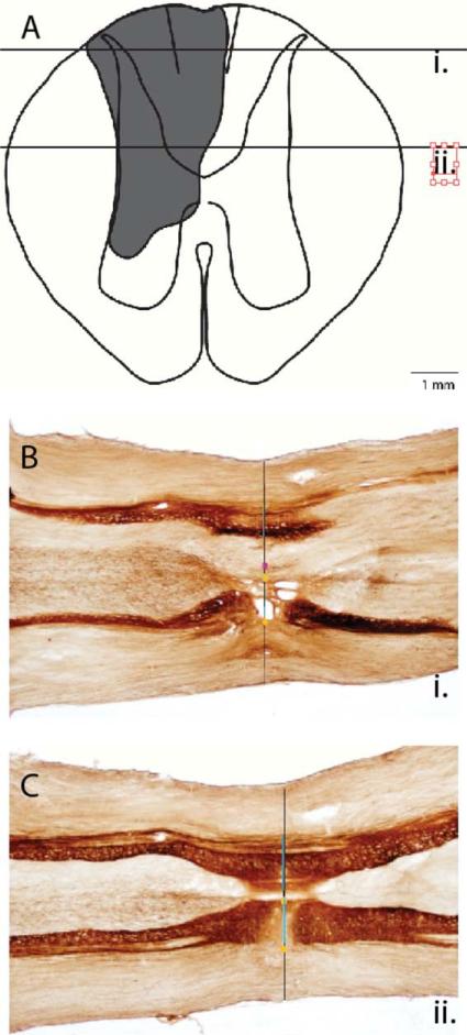
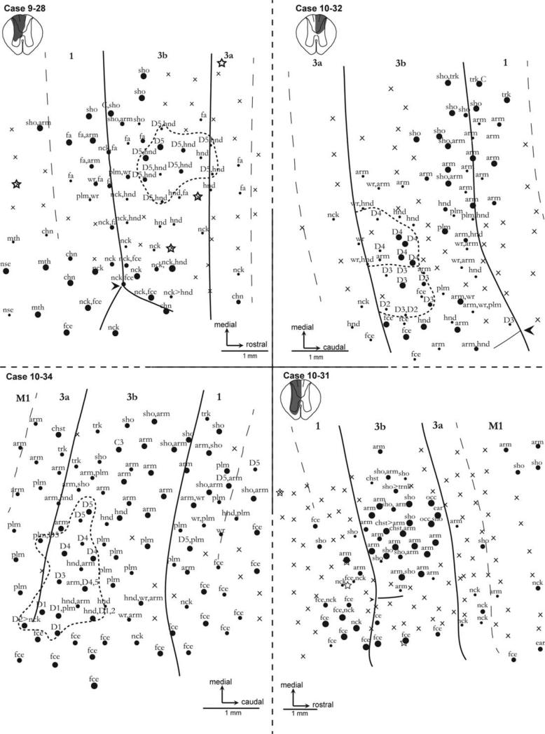
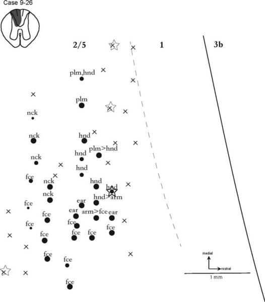
In the present study, we used microelectrode recordings of multiunit responses to evaluate patterns of the reactivation of somatosensory cortex after sensory loss produced by spinal cord lesions in the common marmoset (Callithrix jacchus). These New World monkeys have become a popular model in studies of cortical organization and function. Primary somatosensory cortex and adjoining somatosensory areas can become extensively deactivated by lesions of somatosensory afferents as they ascend in the dorsal columns of the cervical spinal cord. Six to 7 weeks after complete lesions of the cuneate fasciculus subserving the forelimb at cervical levels 5-6, the hand region in contralateral areas 3b and 1 was reactivated by inputs from the forelimb, but excluded representations of some or all digits. In a similar manner, recording sites from the forelimb region of areas 2-5 responded to parts of the forelimb but not to digits after an extensive lesion of the contralateral cuneate fasciculus at C5-C6. Lesions that damaged only the gracile fasciculus or a small percentage of the cuneate fasciculus did not produce changes in the gross hand representation in contralateral areas 3b, 3a, 1, and 2. Finally, a complete but lower lesion of the cuneate fasciculus at C8 produced some abnormalities in the reactivation, but the digits were represented. The results indicate that areas 3a, 3b, 1, and 2-5 of the somatosensory cortex are extensively reactivated after large, apparently complete lesions of the contralateral cuneate fasciculus, but afferents from the digits may not contribute to their reactivation.
Keywords: New World monkey; cortical plasticity; somatosensory.
Copyright © 2013 Wiley Periodicals, Inc.
Figures


Similar articles
-
Spinal cord neuron inputs to the cuneate nucleus that partially survive dorsal column lesions: A pathway that could contribute to recovery after spinal cord injury.J Comp Neurol. 2015 Oct 1;523(14):2138-60. doi: 10.1002/cne.23783. Epub 2015 Jun 2. J Comp Neurol. 2015. PMID: 25845707 Free PMC article.
-
Reorganization of somatosensory cortical areas 3b and 1 after unilateral section of dorsal columns of the spinal cord in squirrel monkeys.J Neurosci. 2011 Sep 21;31(38):13662-75. doi: 10.1523/JNEUROSCI.2366-11.2011. J Neurosci. 2011. PMID: 21940457 Free PMC article.
-
Intracortical connections are altered after long-standing deprivation of dorsal column inputs in the hand region of area 3b in squirrel monkeys.J Comp Neurol. 2016 May 1;524(7):1494-526. doi: 10.1002/cne.23921. Epub 2015 Dec 8. J Comp Neurol. 2016. PMID: 26519356 Free PMC article.
-
Cortical and subcortical plasticity in the brains of humans, primates, and rats after damage to sensory afferents in the dorsal columns of the spinal cord.Exp Neurol. 2008 Feb;209(2):407-16. doi: 10.1016/j.expneurol.2007.06.014. Epub 2007 Jul 6. Exp Neurol. 2008. PMID: 17692844 Free PMC article. Review.
-
The reactivation of somatosensory cortex and behavioral recovery after sensory loss in mature primates.Front Syst Neurosci. 2014 May 12;8:84. doi: 10.3389/fnsys.2014.00084. eCollection 2014. Front Syst Neurosci. 2014. PMID: 24860443 Free PMC article. Review.
Cited by
-
Spinal cord neuron inputs to the cuneate nucleus that partially survive dorsal column lesions: A pathway that could contribute to recovery after spinal cord injury.J Comp Neurol. 2015 Oct 1;523(14):2138-60. doi: 10.1002/cne.23783. Epub 2015 Jun 2. J Comp Neurol. 2015. PMID: 25845707 Free PMC article.
-
Spatiotemporal trajectories of reactivation of somatosensory cortex by direct and secondary pathways after dorsal column lesions in squirrel monkeys.Neuroimage. 2016 Nov 15;142:431-453. doi: 10.1016/j.neuroimage.2016.08.015. Epub 2016 Aug 12. Neuroimage. 2016. PMID: 27523450 Free PMC article.
-
Altered Spatiotemporal Dynamics of Cortical Activation to Tactile Stimuli in Somatosensory Area 3b and Area 1 of Monkeys after Spinal Cord Injury.eNeuro. 2016 Sep 29;3(5):ENEURO.0095-16.2016. doi: 10.1523/ENEURO.0095-16.2016. eCollection 2016 Sep-Oct. eNeuro. 2016. PMID: 27699211 Free PMC article.
-
Common marmoset (Callithrix jacchus) as a primate model for behavioral neuroscience studies.J Neurosci Methods. 2017 Jun 1;284:35-46. doi: 10.1016/j.jneumeth.2017.04.004. Epub 2017 Apr 8. J Neurosci Methods. 2017. PMID: 28400103 Free PMC article.
-
Comparative Functional Anatomy of Marmoset Brains.ILAR J. 2020 Dec 31;61(2-3):260-273. doi: 10.1093/ilar/ilaa026. ILAR J. 2020. PMID: 33550381 Free PMC article.
References
-
- Bennett GJ, Seltzer Z, Lu GW, Nishikawa N, Dubner R. The cells of origin of the dorsal column postsynaptic projection in the lumbosacral enlargements of cats and monkeys. Somatosens Res. 1983;1:131–149. - PubMed
-
- Carlson M, Huerta MF, Cusick CG, Kaas JH. Studies on the evolution of multiple somatosensory representations in primates: the organization of anterior parietal cortex in the New World Callitrichid, Saguinus. J Comp Neurol. 1986;246:409–426. - PubMed
-
- Darian-Smith C, Brown S. Functional changes at periphery and cortex following dorsal root lesions in adult monkeys. Nat Neurosci. 2000;3:476–481. - PubMed
Publication types
MeSH terms
Substances
Grants and funding
LinkOut - more resources
Full Text Sources
Other Literature Sources
Medical
Miscellaneous
