. 2013 Jun 18;51(1):99-108.
doi: 10.1530/JME-12-0251. Print 2013.

Regulation of the osterix (Osx, Sp7) promoter by osterix and its inhibition by parathyroid hormone

Affiliations

Regulation of the osterix (Osx, Sp7) promoter by osterix and its inhibition by parathyroid hormone

Richard Barbuto et al. J Mol Endocrinol. .

Abstract

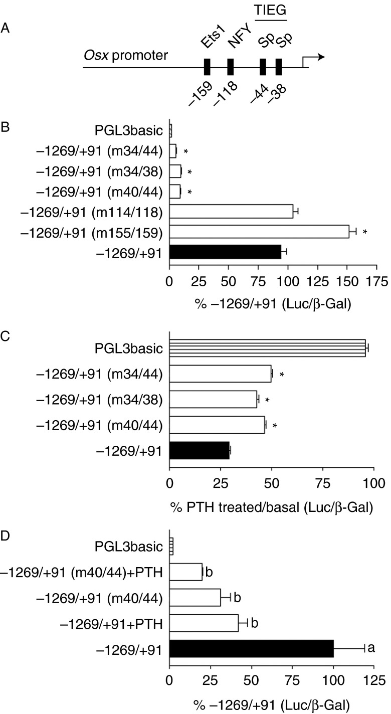
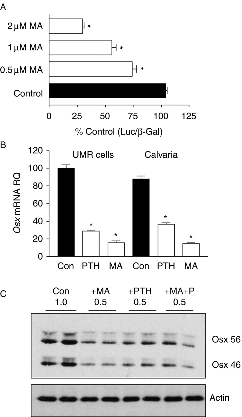
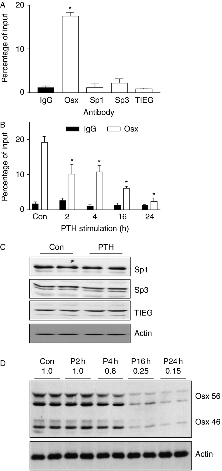
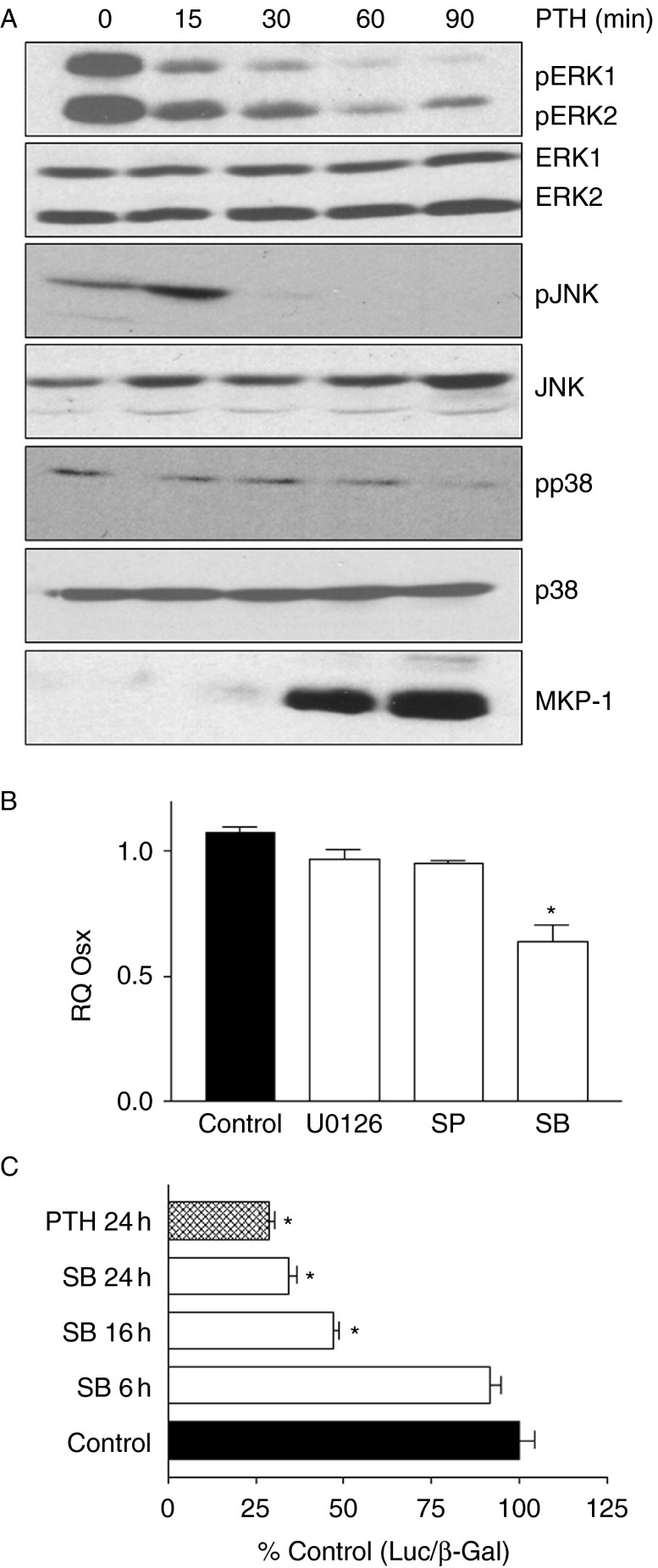
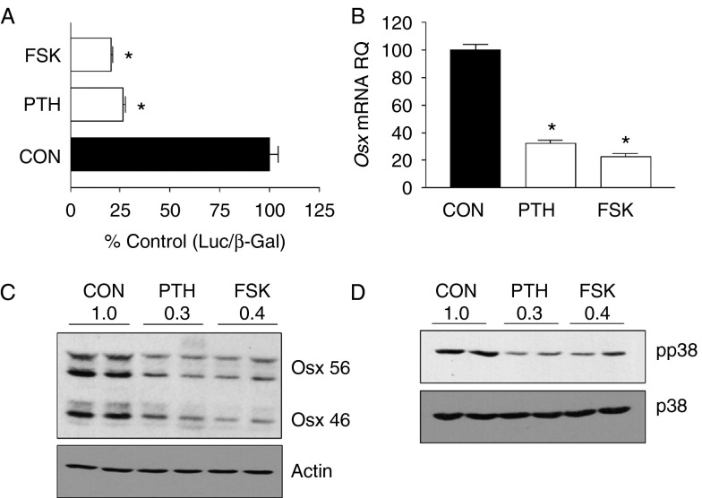
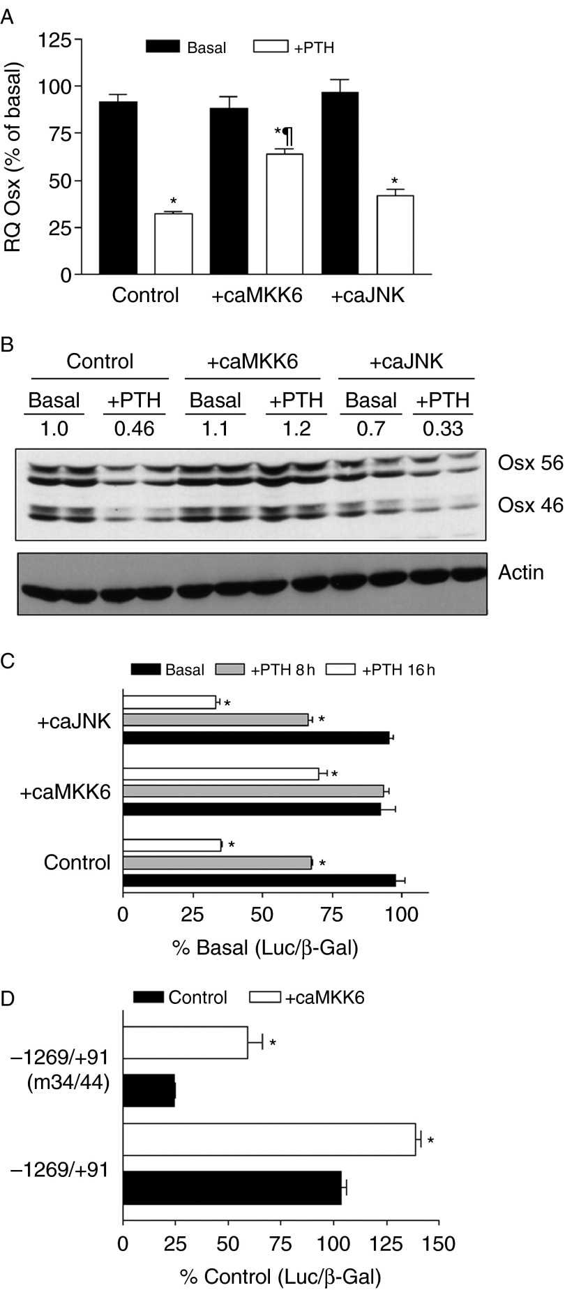
Osterix (Osx, Sp7) is a zinc-finger transcription factor belonging to the specificity protein (Sp) family expressed in cells of the osteoblast lineage in the developing skeleton where it regulates expression of a number of osteoblastic genes. We previously reported inhibition of osterix mRNA and protein by parathyroid hormone (PTH) stimulation of cAMP in osteoblasts. We here show that Osx expression in osteoblasts is regulated by Sp proteins as demonstrated by mithramycin A inhibition of Osx mRNA and OSX protein levels. Mutation of putative transcription factor binding sites within the Osx promoter demonstrated a tandem repeat sequence that selectively binds OSX but not other Sp factors expressed in osteoblasts (Sp1, Sp3, or Tieg (Klf10)). Mutation of either or both the repeat sequences inhibited 90% of the promoter activity and also abrogated some of the PTH-mediated inhibition of the promoter. Previous studies have shown growth factor regulation of Osx expression by MAPK proteins, particularly p38 phosphorylation of OSX that increases its transcriptional activity. PTH stimulation of osteoblasts inhibits MAPK components (ERK, JNK, and p38) but inhibition of Osx mRNA and protein expression by PTH was selectively mimicked by p38 inhibition and expression of constitutively active MKK6, which stimulates p38, blocked PTH inhibition of OSX. Together, our studies suggest that OSX autoregulation is a major mechanism in osteoblasts and that PTH stimulation inhibits osterix by inhibition of p38 MAPK regulation of OSX.

Keywords: Sp factor; autoregulation; osteoblasts; osterix; parathyroid hormone.


Figures

Figure 1
Figure 1
Mutational analysis of the Osx promoter. (A) Schematic representation of the relative positions of putative transcription factor binding sites in the −1269/+91 Osx promoter. (B) UMR106-01 cells transfected with the −1269/+91 Osx promoter reporter; promoter constructs with mutations in the regions indicated in brackets or empty vector (PGL3basic). Cells were assayed for luciferase and β-galactosidase activities 24 h after transfection. Corrected luciferase activities expressed relative to the wild-type promoter are shown. (C) PTH regulation of the indicated promoter luciferase constructs or empty vector was assessed using 10 nM rPTH(1–34) for 16 h before measuring luciferase activity. The values shown indicate luciferase activity in the presence of PTH relative to that in the basal state. (D) Luciferase activity of −1269/+91 Osx promoter reporter and the mutation of the first Sp site (−40/−44) reporter were tested for basal and PTH-regulated activity in primary mouse osteoblasts. Cells were treated with or without 10 nM rPTH(1–34) for 16 h before measuring luciferase activity. The values shown indicate luciferase activity in the presence of PTH relative to that in the basal state, cells transfected with empty vector were assessed without PTH stimulation. Statistical significance in B and C: *P<0.001 relative to −1269/+91; in D, statistical differences between bars are indicated by differences in letters P<0.05.
Figure 2
Figure 2
Mithramycin A (MA) inhibits Osx promoter activity and OSX expression. (A) UMR106-01 cells were transfected with −1269/+91 Osx promoter and incubated in the presence of increasing concentrations of MA or vehicle control for 8 h before assessment of luciferase activity corrected for β-galactosidase activity in the same cells. (B) UMR106-01 cells or neonatal mouse calvaria were incubated in the presence of 10 nM rPTH(1–34) (PTH), 2 μM MA (MA), or vehicle control (Con) for 16 h before extraction of RNA and assessment of Osx mRNA levels by real-time PCR relative to the levels of GAPDH in each sample. Bars indicate Osx mRNA levels relative to Con for each cell/tissue type. Statistical significance in A and B: *P<0.001 relative to control values. (C) The effect of incubation of UMR106-01 cells with 1 μM MA (+MA), 10 nM rPTH(1–34) (+PTH), 1 μM MA and 10 nM rPTH(1–34) (+MA+P) or vehicle control (Con) for 16 h on OSX and α-actin protein expression were assessed by SDS–PAGE and immunoblot. Two isoforms of OSX were expressed and both were suppressed by MA or PTH by 50%. Duplicate samples were assessed in each experiment and each experiment was repeated twice with representative blots shown.
Figure 3
Figure 3
Osterix but not other Sp factors bind to the Osx promoter. (A) Chromatin prepared from UMR106-01 cells was assessed for transcription factor binding to the Osx promoter proximal to the two Sp binding sites (−44/−34). Following immunoprecipitation with antibodies specific to the indicated transcription factors, the amount of Osx promoter binding to each factor was assessed by real-time PCR. Bars indicate the amount of Osx DNA in each sample relative to the total input amount in the assay. Nonspecific binding was assessed using IgG in the immunoprecipitation assay. *Statistical significance: P<0.0001 relative to IgG. (B) UMR106-01 cells were incubated with 10 nM rPTH(1–34) for the indicated times before cell extraction and chromatin preparation. PTH stimulation significantly decreased OSX binding to its own promoter within 2 h of stimulation and this continued to decrease over 24 h. Statistical significance: *P<0.05 relative to control values. (C) Immunoblots of 25 μg samples of UMR106-01 cell extracts showing expression of each of the transcription factors assessed in the ChIP assays. Incubation of the cells with 10 nM rPTH(1–34) for 8 h had no effect on the levels of any of these proteins. (D) Immunoblots of OSX expression in UMR106-01 cells treated with 10 nM rPTH(1–34) over the same time periods as used for ChIP assays show that OSX protein was decreased with treatments of 4 h or longer.
Figure 4
Figure 4
Effect of PTH stimulation on the levels of MAPKs and the effect of MAPK inhibitors on Osx mRNA and Osx promoter luciferase activity. (A) UMR106-01 cells were incubated for the indicated times with 10 nM rPTH(1–34) before extraction of cellular proteins. 10 μg samples of cellular extracts were analyzed by SDS–PAGE and immunoblots using specific antibodies to each of the indicated MAPKs and their activated (phosphorylated) forms as shown on the side of each blot. Levels of MKP-1, a phosphatase that can dephosphorylate MAPKs, are also shown in the bottom panel. (B) UMR106-01 cells were incubated with 10 μM concentrations of U0126, a specific inhibitor of ERK1/2; SP600125, a specific inhibitor of JNK (SP); SB203580, a specific inhibitor of p38 MAPK (SB), or vehicle control for 16 h before extraction of cellular RNA and assessment of Osx mRNA by real-time PCR. (C) UMR106-01 cells were transfected with −1269/+91 promoter luciferase construct and the cells were then incubated with either 10 μM SB203580 for the indicated times or 10 nM rPTH(1–34) for 24 h before assessment of luciferase activity. Statistical significance in B and C indicated by *P<0.05 relative to control values.
Figure 5
Figure 5
Effects of PTH on OSX and p38MAPK are also seen with forskolin stimulation of UMR cells. (A) UMR106-01 cells were transfected with the −1269/+91 Osx promoter reporter construct and treated with 10 nM rPTH(1–34), 10 μM forskolin, or vehicle control for 16 h before measuring luciferase activity. The values shown indicate luciferase activity relative to that in the basal state. (B) UMR106-01 cells were incubated in the presence of 10 nM rPTH(1–34) (PTH), 10 μM forskolin (FSK), or vehicle control (Con) for 16 h before extraction of RNA and assessment of Osx mRNA levels by real-time PCR relative to the levels of GAPDH in each sample. Bars indicate Osx mRNA levels relative to Con for each cell/tissue type. Statistical significance in A and B *P<0.001 relative to control values. (C) UMR106-01 cells were incubated in the presence of 10 nM rPTH(1–34) (PTH), 10 μM forskolin (FSK), or vehicle control (Con) for 16 h before extraction of protein and assessment of OSX protein levels by immunoblot relative to the levels of actin in each sample. (D) UMR106-01 cells were incubated for the 90 min with 10 nM rPTH(1–34) (PTH), 10 μM forskolin (FSK), or vehicle control (Con) before extraction of cellular proteins. Samples of cellular extracts were analyzed by SDS–PAGE and immunoblots using specific antibodies to the indicated p38MAPK and its activated form (pp38) as shown on the side of each blot.
Figure 6
Figure 6
Expression of constitutively active MKK6 but not JNK abrogates PTH inhibition of Osx mRNA, protein, and Osx promoter activity. (A) UMR106-01 cells were transfected with cDNAs encoding caMKK6, an upstream kinase that activated p38 MAPK or JNKK2-JNK1, a constitutively active JNK1. The following day, cells were incubated with or without 10 nM rPTH(1–34) for 8 h before RNA extraction and assessment of Osx mRNA by real-time PCR. *Values significantly different from basal levels, P<0.05; values significantly different from control +PTH, P<0.05. (C and D) UMR106-01 cells were transfected with caMKK6, JNKK2-JNK1 (caJNK), or a control cDNA (control) in addition to −1269/+91 promoter luciferase construct. Cells were then incubated with 10 nM rPTH(1–34) for 8 or 16 h and compared with cells incubated without PTH (Basal). Luciferase activities relative to control basal are plotted. *Values significantly different from control basal levels, P<0.01. (B) UMR106-01 cells were transfected with caMKK6, JNKK2-JNK1 (caJNK), or a control cDNA (control) and then incubated with 10 nM rPTH(1–34) for 8 h before extraction of cellular proteins. 25 μg samples of protein extracts were run on SDS–PAGE followed by immunoblot for OSX or actin. The numbers on top of the blots indicate the levels of OSX protein relative to control basal levels.

References

    1. Baek WY, Lee MA, Jung JW, Kim SY, Akiyama H, de Crombrugghe B, Kim JE. Positive regulation of adult bone formation by osteoblast-specific transcription factor osterix. Journal of Bone and Mineral Research. 2009;24:1055–1065. doi: 10.1359/jbmr.081248. - DOI - PMC - PubMed
    1. Bianchi EN, Ferrari SL. β-Arrestin2 regulates parathyroid hormone effects on a p38 MAPK and NFkappaB gene expression network in osteoblasts. Bone. 2009;45:716–725. doi: 10.1016/j.bone.2009.06.020. - DOI - PMC - PubMed
    1. Blume SW, Snyder RC, Ray R, Thomas S, Koller CA, Miller DM. Mithramycin inhibits SP1 binding and selectively inhibits transcriptional activity of the dihydrofolate reductase gene in vitro and in vivo. Journal of Clinical Investigation. 1991;88:1613–1621. doi: 10.1172/JCI115474. - DOI - PMC - PubMed
    1. Celil AB, Campbell PG. BMP-2 and insulin-like growth factor-I mediate Osterix (Osx) expression in human mesenchymal stem cells via the MAPK and protein kinase D signaling pathways. Journal of Biological Chemistry. 2005;280:31353–31359. doi: 10.1074/jbc.M503845200. - DOI - PubMed
    1. Cuadrado A, Nebreda AR. Mechanisms and functions of p38 MAPK signalling. Biochemical Journal. 2010;429:403–417. doi: 10.1042/BJ20100323. - DOI - PubMed

Publication types

MeSH terms