

A case of hemophagocytic syndrome in a patient with fulminant ulcerative colitis superinfected by cytomegalovirus
- PMID: 23682230
- PMCID: PMC3654134
- DOI: 10.3904/kjim.2013.28.3.352
A case of hemophagocytic syndrome in a patient with fulminant ulcerative colitis superinfected by cytomegalovirus
Abstract
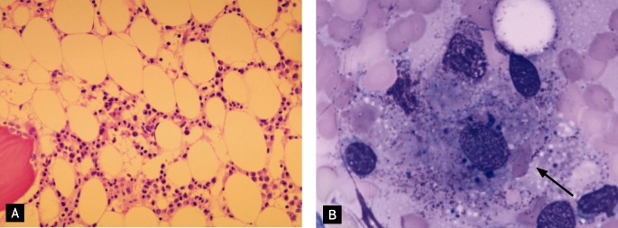
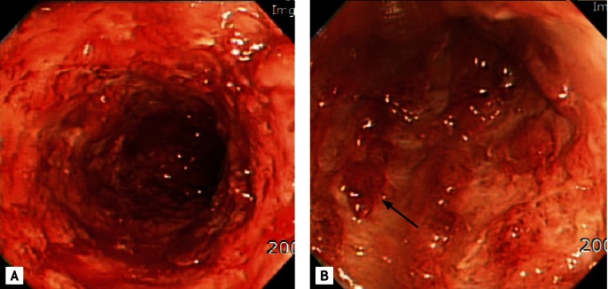
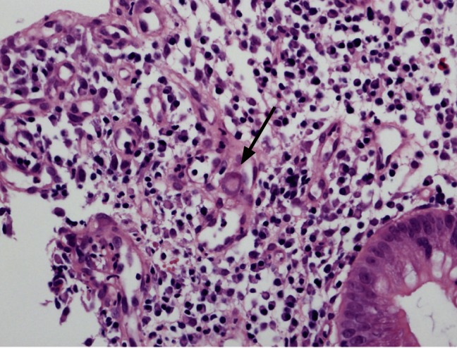
Hemophagocytic syndrome (HPS) is an uncommon hematological disorder that manifests as fever, splenomegaly, and jaundice, with hemophagocytosis in the bone marrow and other tissues pathologically. Secondary HPS is associated with malignancy and infection, especially viral infection. The prevalence of cytomegalovirus (CMV) infection in ulcerative colitis (UC) patients is approximately 16%. Nevertheless, HPS in UC superinfected by CMV is very rare. A 52-year-old female visited the hospital complaining of abdominal pain and hematochezia for 6 days. She was diagnosed with UC 3 years earlier and had been treated with sulfasalazine, but had stopped her medication 4 months earlier. On admission, her spleen was enlarged. The peripheral blood count revealed pancytopenia and bone marrow aspiration smears showed hemophagocytosis. Viral studies revealed CMV infection. She was treated successfully with ganciclovir. We report this case with a review of the related literature.
Keywords: Colitis, ulcerative; Cytomegalovirus infections; Inflammatory bowel diseases; Lymphohistiocytosis, hemophagocytic.
Conflict of interest statement
No potential conflict of interest relevant to this article is reported.
Figures
Similar articles
-
[A case of hemophagocytic syndrome in an ulcerative colitis patient].Korean J Gastroenterol. 2010 Jul;56(1):45-8. doi: 10.4166/kjg.2010.56.1.45. Korean J Gastroenterol. 2010. PMID: 20695130 Korean.
-
[Acute cytomegalovirus infection-associated hemophagocytic syndrome in a patient treated with azathioprine].Orv Hetil. 2013 Dec 8;154(49):1959-61. doi: 10.1556/OH.2013.29764. Orv Hetil. 2013. PMID: 24292114 Hungarian.
-
Hemophagocytic syndrome caused by fulminant ulcerative colitis and cytomegalovirus infection: report of a case.Dis Colon Rectum. 2004 Jul;47(7):1250-3; discussion 1253-5. doi: 10.1007/s10350-004-0543-x. Epub 2004 May 19. Dis Colon Rectum. 2004. PMID: 15148644
-
Toxic megacolon associated with cytomegalovirus infection in a patient with steroid-naïve ulcerative colitis.Intern Med. 2012;51(19):2739-43. doi: 10.2169/internalmedicine.51.8145. Epub 2012 Oct 1. Intern Med. 2012. PMID: 23037465 Review.
-
Cytomegalovirus and ulcerative colitis: Place of antiviral therapy.World J Gastroenterol. 2016 Feb 14;22(6):2030-45. doi: 10.3748/wjg.v22.i6.2030. World J Gastroenterol. 2016. PMID: 26877608 Free PMC article. Review.
Cited by
-
Approach to cytomegalovirus infections in patients with ulcerative colitis.Korean J Intern Med. 2017 May;32(3):383-392. doi: 10.3904/kjim.2017.087. Epub 2017 Apr 20. Korean J Intern Med. 2017. PMID: 28490715 Free PMC article. Review.
-
Haemophagocytic lymphohistiocytosis in inflammatory bowel disease with virus infection.Prz Gastroenterol. 2015;10(2):78-82. doi: 10.5114/pg.2015.48995. Epub 2015 Oct 2. Prz Gastroenterol. 2015. PMID: 26557937 Free PMC article. Review.
-
Hemophagocytic Lymphohistiocytosis and Inflammatory Bowel Disease: Case Report and Systematic Review.Inflamm Intest Dis. 2020 Jun;5(2):49-58. doi: 10.1159/000506514. Epub 2020 Mar 10. Inflamm Intest Dis. 2020. PMID: 32596254 Free PMC article. Review.
-
Concomitant Hemophagocytic Lymphohistiocytosis and Cytomegalovirus Disease: A Case Based Systemic Review.Front Med (Lausanne). 2022 Apr 19;9:819465. doi: 10.3389/fmed.2022.819465. eCollection 2022. Front Med (Lausanne). 2022. PMID: 35514747 Free PMC article.
-
Secondary Hemophagocytosis in Propionic Acidemia.Iran J Pediatr. 2015 Jun;25(3):e339. doi: 10.5812/ijp.25(3)2015.339. Epub 2015 Jun 27. Iran J Pediatr. 2015. PMID: 26199705 Free PMC article. No abstract available.
References
-
- James DG, Stone CD, Wang HL, Stenson WF. Reactive hemophagocytic syndrome complicating the treatment of inflammatory bowel disease. Inflamm Bowel Dis. 2006;12:573–580. - PubMed
-
- Janka GE. Hemophagocytic syndromes. Blood Rev. 2007;21:245–253. - PubMed
-
- Kim JJ, Simpson N, Klipfel N, Debose R, Barr N, Laine L. Cytomegalovirus infection in patients with active inflammatory bowel disease. Dig Dis Sci. 2010;55:1059–1065. - PubMed
-
- Koketsu S, Watanabe T, Hori N, Umetani N, Takazawa Y, Nagawa H. Hemophagocytic syndrome caused by fulminant ulcerative colitis and cytomegalovirus infection: report of a case. Dis Colon Rectum. 2004;47:1250–1253. - PubMed
Publication types
MeSH terms
Substances
LinkOut - more resources
Full Text Sources
Other Literature Sources
Medical