

Comment
doi: 10.7554/eLife.00804.
Make or break for mitochondria
Affiliations
- PMID: 23682317
- PMCID: PMC3654434
- DOI: 10.7554/eLife.00804
Item in Clipboard
Comment
Make or break for mitochondria
Elife.
.
Abstract
Ensuring that mitochondrial DNA is successfully divided between daughter mitochondria involves a complex series of interactions with the endoplasmic reticulum and a variety of enzymes.
Keywords: ERMES; Gem1; Miro; S. cerevisiae; mitochondria; mitochondrial DNA.
Conflict of interest statement
Figures
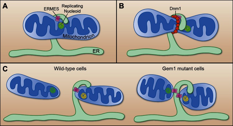
(A) Murley et al. demonstrate that the ERMES tethering complex (pink) localizes to the region where the endoplasmic reticulum (ER) makes contact with a mitochondrion in yeast, and where the nucleoid that contains the mitochondrial DNA is replicating. (B) The mitochondrion undergoes constriction at this contact site, which allows a helix of Dnm1 (red) to form around it. (C) This helix leads to further constriction and, ultimately, to the division of the mitochondrion and the formation of two daughter mitochondria in wild-type cells (left). Each daughter mitochondrion has its own nucleoid. Significantly, one daughter remains attached to the ER via ERMES, which remains intact through the division process. However, in cells lacking Gem1, both of the daughter mitochondria remain tethered to the same ER segment, and both have somewhat unusual shapes (right).
Comment on
-
ER-associated mitochondrial division links the distribution of mitochondria and mitochondrial DNA in yeast.Elife. 2013 May 14;2:e00422. doi: 10.7554/eLife.00422. Elife. 2013. PMID: 23682313 Free PMC article.
References
Publication types
MeSH terms
Substances
LinkOut - more resources
Full Text Sources
Other Literature Sources
Molecular Biology Databases
