. 2012;2(2):1000107.
doi: 10.4172/2161-1025.1000107.

Quantitative and Sensitive Detection of Cancer Genome Amplifications from Formalin Fixed Paraffin Embedded Tumors with Droplet Digital PCR

Affiliations

Quantitative and Sensitive Detection of Cancer Genome Amplifications from Formalin Fixed Paraffin Embedded Tumors with Droplet Digital PCR

Lincoln Nadauld et al. Transl Med (Sunnyvale). 2012.

Abstract

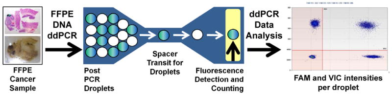
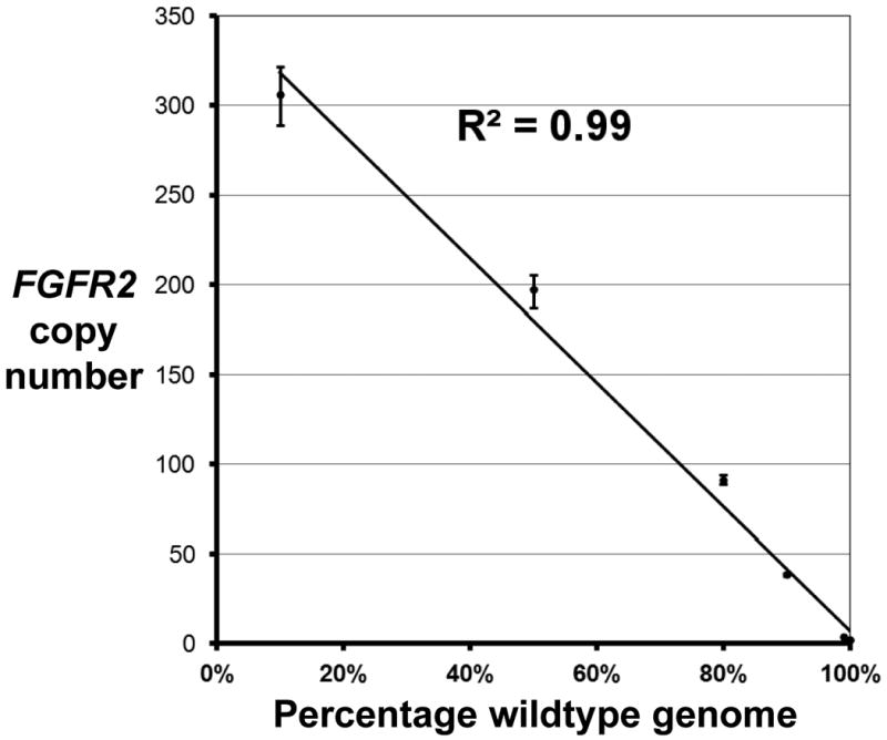
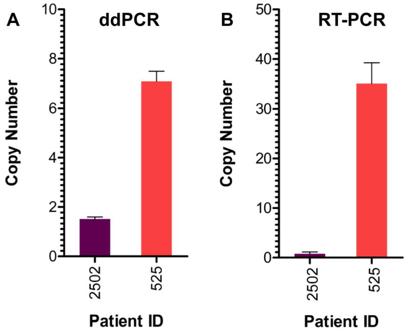
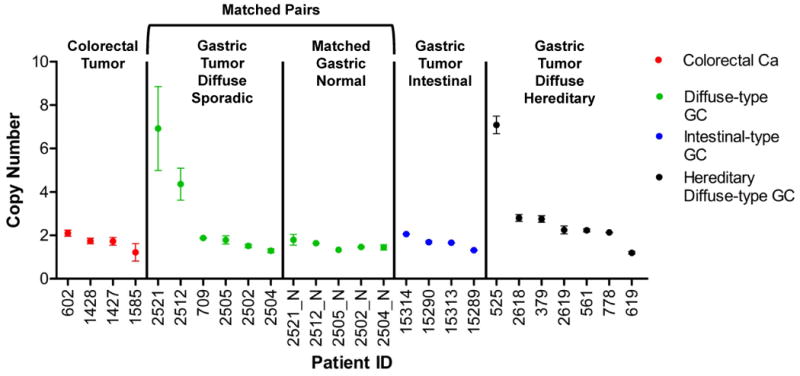
For the analysis of cancer, there is great interest in rapid and accurate detection of cancer genome amplifications containing oncogenes that are potential therapeutic targets. The vast majority of cancer tissue samples are formalin fixed and paraffin embedded (FFPE) which enables histopathological examination and long term archiving. However, FFPE cancer genomic DNA is oftentimes degraded and generally a poor substrate for many molecular biology assays. To overcome the issues of poor DNA quality from FFPE samples and detect oncogenic copy number amplifications with high accuracy and sensitivity, we developed a novel approach. Our assay requires nanogram amounts of genomic DNA, thus facilitating study of small amounts of clinical samples. Using droplet digital PCR (ddPCR), we can determine the relative copy number of specific genomic loci even in the presence of intermingled normal tissue. We used a control dilution series to determine the limits of detection for the ddPCR assay and report its improved sensitivity on minimal amounts of DNA compared to standard real-time PCR. To develop this approach, we designed an assay for the fibroblast growth factor receptor 2 gene (FGFR2) that is amplified in a gastric and breast cancers as well as others. We successfully utilized ddPCR to ascertain FGFR2 amplifications from FFPE-preserved gastrointestinal adenocarcinomas.

Keywords: archival cancer samples; cancer; copy number variations; gene amplifications.


Figures

Figure 1
Figure 1
General workflow of droplet digital PCR in amplification analysis of archival cancer samples. DNA is extracted from an archival cancer sample. An example of a formalin-fixed paraffin block of gastric adencarcinoma with an accompanying stained section is shown. After DNA extraction, droplet PCR is conducted with a specific set of PCR primers and fluorescent probe. Post-PCR emulsion droplets are streamed single-file into a capillary that leads past a two-color detector; where the positive droplets for the target and reference genes are counted for copy number quantitation with two different dyes such as 6-FAM and VIC. One dye is specific to the control loci and the other to the loci being measured.
Figure 2
Figure 2
Measurement of FGFR2 copy number from a dilution series of tumor versus normal genomic DNA using ddPCR. To ascertain the limits of quantifying amplifications, KatoIII genomic DNA, known to harbor FGFR2 amplification, was diluted with a normal diploid genomic DNA in decreasing ratios. Data points with error bars represent the drop in FGFR2 copy number as the KatoIII genomic DNA is diluted with normal genomic DNA.
Figure 3
Figure 3
Comparison of ddPCR versus real-time PCR for measuring FGFR2 copy number in FFPE tissue. FGFR2 copy number was measured in two different patient samples by ddPCR (panel A) and real-time PCR (panel B). The FFPE sample from Patient 2502 (purple bars) represents normal gastric tissue. The FFPE sample from Patient 525 (red bars) is a gastric adenocarcinoma sample. Data are plotted as copy number per diploid genome.
Figure 4
Figure 4
FGFR2 copy number measured by ddPCR in archival FFPE cancer samples. Copy number of the FGFR2 gene were measured in human colon carcinomas (red dots, n=4), diffuse-type gastric carcinomas (green dots, n=6), intestinal-type gastric carcinomas (blue dots, n=4), and hereditary diffuse gastric cancers (black dots, n=7). Patient 525 is a positive control for the FGFR2 amplification. The X-axis numbers indicate patient identification numbers; “N” indicates a matched normal tissue control for that patient identification number. Error bars represent standard deviation of a minimum of 3 experimental replicates.

References

    1. Weinstein IB, Joe A. Oncogene addiction. Cancer Res. 2008;68:3077–3080. discussion 3080. - PubMed
    1. Settleman J. Oncogene addiction. Curr Biol. 2012;22:R43–44. - PubMed
    1. Kallioniemi OP, Kallioniemi A, Kurisu W, Thor A, Chen LC, Smith HS, Waldman FM, Pinkel D, Gray JW. ERBB2 amplification in breast cancer analyzed by fluorescence in situ hybridization. Proc Natl Acad Sci U S A. 1992;89:5321–5325. - PMC - PubMed
    1. Dressman D, Yan H, Traverso G, Kinzler KW, Vogelstein B. Transforming single DNA molecules into fluorescent magnetic particles for detection and enumeration of genetic variations. Proceedings of the National Academy of Sciences of the United States of America. 2003;100:8817–8822. - PMC - PubMed
    1. Hindson BJ, Ness KD, Masquelier DA, Belgrader P, Heredia NJ, Makarewicz AJ, Bright IJ, Lucero MY, Hiddessen AL, Legler TC, et al. High-throughput droplet digital PCR system for absolute quantitation of DNA copy number. Anal Chem. 2011;83:8604–8610. - PMC - PubMed

LinkOut - more resources