Review
. 2013 Jul 8:36:165-82.
doi: 10.1146/annurev-neuro-062012-170249. Epub 2013 May 15.

Superior colliculus and visual spatial attention

Affiliations
Review

Superior colliculus and visual spatial attention

Richard J Krauzlis et al. Annu Rev Neurosci. .

Abstract

The superior colliculus (SC) has long been known to be part of the network of brain areas involved in spatial attention, but recent findings have dramatically refined our understanding of its functional role. The SC both implements the motor consequences of attention and plays a crucial role in the process of target selection that precedes movement. Moreover, even in the absence of overt orienting movements, SC activity is related to shifts of covert attention and is necessary for the normal control of spatial attention during perceptual judgments. The neuronal circuits that link the SC to spatial attention may include attention-related areas of the cerebral cortex, but recent results show that the SC's contribution involves mechanisms that operate independently of the established signatures of attention in visual cortex. These findings raise new issues and suggest novel possibilities for understanding the brain mechanisms that enable spatial attention.


Figures

Figure 1
Figure 1
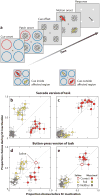
Effects of superior colliculus (SC) inactivation on pursuit target selection. (a) The task was to smoothly track the target defined by a precue. Performance on this two-alternative task was measured before and after SC inactivation. (b) The area affected by SC inactivation was estimated by measuring the change in peak velocity of saccades to visual targets. (c) Changes in performance when the target appeared inside the affected part of the visual field. The distributions of target choices are illustrated by plotting the mean horizontal eye velocity against mean vertical eye velocity over the first 100 ms of the pursuit eye movement response. SC inactivation reduced correct choices from 66% to 15% correct. (d) When the distracter appeared in the affected part of the visual field, correct pursuit choices improved from 71% correct to 96% correct. Overall, SC inactivation biased choices in favor of the stimulus appearing outside the affected region, even though these required movements toward the affected field. Adapted from Nummela & Krauzlis (2010).
Figure 2
Figure 2
Changes in superior colliculus (SC) activity during covert shifts of spatial attention. The sample visual neuron showed enhanced activity (shaded area) when the animal was shown a spatially precise cue (b) compared to when the animal was shown no cue (a). (cd) Sample visual-motor neuron under the same task conditions. Both the visual and visual-motor neurons responded to the presentation of the “C” (blue), but this activity was higher when the location of the “C” was cued than when it was not cued. Also, the visual-motor neuron showed activity during the time epoch before the presentation of the “C” [attention shift period (ASP), orange], but the visual neuron did not. Abbreviations: F, fixation spot; RF, receptive field of neuron; T, choice target. Adapted from Ignashchenkova et al. (2004).
Figure 3
Figure 3
Impairments in covert selection of signals for perceptual judgments during superior colliculus (SC) inactivation. (a) Task design. A color cue indicated the relevant motion patch, which contained a brief pulse of motion in one of four possible directions. The animal indicated its choice either by making a saccade in the discriminated direction or by pressing a corresponding button. (b) When the cued signal was in the affected region, animals ignored this signal and instead based their choices on the foil. (c) Conversely, when the foil signal appeared in the affected region, subjects tended to ignore the foil. (de) Similar results were obtained in the absence of saccades during a button-press version of the task. Filled symbols show data from muscimol injection experiments; open symbols, from saline control injections. Adapted from Lovejoy & Krauzlis (2010).
Figure 4
Figure 4
Neuronal correlates of spatial attention remain intact in visual cortex during superior colliculus (SC) inactivation despite behavioral deficits in attention. (a) Task design. A cue indicated the relevant motion patch, and later either this cued patch or the foil patch changed its direction. If the cued patch changed, the animals reported this detection by pressing a button. (bc) Neuronal activity was recorded in the medial superior temporal area (MST) before (b) and during (c) SC inactivation. (de) Activity of a sample neuron during the attention task before (d) and during (e) SC inactivation. Despite large deficits in task performance caused by SC inactivation, the modulation of neuronal activity by spatial cues remained intact. (f–g) Receptive field and tuning properties before (f) and during (g) SC inactivation. The blue shading shows the affected region. Abbreviations: RF, receptive field. Adapted from Zénon & Krauzlis (2012).
Figure 5
Figure 5
Possible ascending neuronal circuits accounting for the superior colliculus (SC) role in spatial attention. Three circuits are outlined in these schematic sections of the monkey brain. One pathway (yellow) passes through the inferior pulvinar (Pi) to the middle temporal area (MT). A second pathway (pink) involves the thalamic reticular nucleus (TRN), which connects to the lateral geniculate nucleus (LGN), which, in turn, projects to the visual cortex (V1 and V2). A third pathway (light blue) passes through the lateral pulvinar (Pl) to the lateral intraparietal area (LIP) and through the mediodorsal thalamus (MD) to the frontal eye fields (FEF).
Figure 6
Figure 6
Possible subcortical neuronal circuits accounting for the superior colliculus (SC) role in spatial attention. One pathway (light blue) passes through the centromedian parafascicular complex (CM-Pf) to the caudate nucleus (Cd) in the basal ganglia. A second pathway (pink) projects to the substantia nigra pars compacta (SNpc). A third circuit (green) involves cholinergic inputs from the pedunculopontine tegmental nucleus (PPTg) and parabigeminal nucleus (PBG). A fourth pathway (yellow) reaches the pontine nuclei (Pn), which relay cortical signals to the cerebellum (Cblm).

References

    1. Aizawa H, Kobayashi Y, Yamamoto M, Isa T. Injection of nicotine into the superior colliculus facilitates occurrence of express saccades in monkeys. J Neurophysiol. 1999;82(3):1642–46. - PubMed
    1. Basso MA, Wurtz RH. Modulation of neuronal activity in superior colliculus by changes in target probability. J Neurosci. 1998;18(18):7519–34. - PMC - PubMed
    1. Bell AH, Fecteau JH, Munoz DP. Using auditory and visual stimuli to investigate the behavioral and neuronal consequences of reflexive covert orienting. J Neurophysiol. 2004;91(5):2172–84. - PubMed
    1. Benevento LA, Port JD. Single neurons with both form/color differential responses and saccade-related responses in the nonretinotopic pulvinar of the behaving macaque monkey. Vis Neurosci. 1995;12(3):523–44. - PubMed
    1. Berman RA, Wurtz RH. Functional identification of a pulvinar path from superior colliculus to cortical area MT. J Neurosci. 2010;30(18):6342–54. - PMC - PubMed

LinkOut - more resources