

From atomic structures to neuronal functions of g protein-coupled receptors
- PMID: 23682660
- PMCID: PMC4594856
- DOI: 10.1146/annurev-neuro-062012-170313
From atomic structures to neuronal functions of g protein-coupled receptors
Abstract
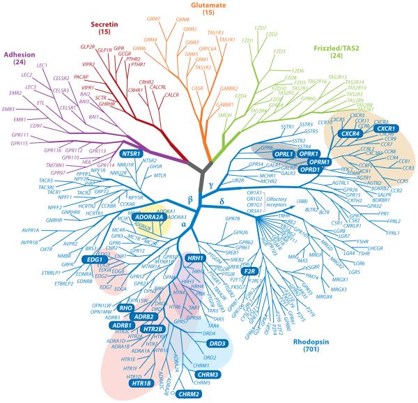
G protein-coupled receptors (GPCRs) are essential mediators of signal transduction, neurotransmission, ion channel regulation, and other cellular events. GPCRs are activated by diverse stimuli, including light, enzymatic processing of their N-termini, and binding of proteins, peptides, or small molecules such as neurotransmitters. GPCR dysfunction caused by receptor mutations and environmental challenges contributes to many neurological diseases. Moreover, modern genetic technology has helped identify a rich array of mono- and multigenic defects in humans and animal models that connect such receptor dysfunction with disease affecting neuronal function. The visual system is especially suited to investigate GPCR structure and function because advanced imaging techniques permit structural studies of photoreceptor neurons at both macro and molecular levels that, together with biochemical and physiological assessment in animal models, provide a more complete understanding of GPCR signaling.
Figures


References
-
- Altier C, Zamponi GW. Analysis of GPCR/ion channel interactions. Methods Mol. Biol. 2011;756:215–25. - PubMed
-
- Anderson DH, Fisher SK. The relationship of primate foveal cones to the pigment epithelium. J. Ultrastruct. Res. 1979;67:23–32. - PubMed
-
- Anderson DH, Fisher SK, Erickson PA, Tabor GA. Rod and cone disc shedding in the rhesus monkey retina: a quantitative study. Exp. Eye Res. 1980;30:559–74. - PubMed
-
- Anderson DH, Fisher SK, Steinberg RH. Mammalian cones: disc shedding, phagocytosis, and renewal. Investig. Ophthalmol. Vis. Sci. 1978;17:117–33. - PubMed
Publication types
MeSH terms
Substances
Grants and funding
LinkOut - more resources
Full Text Sources
Other Literature Sources
