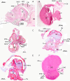
External and internal shell formation in the ramshorn snail Marisa cornuarietis are extremes in a continuum of gradual variation in development
- PMID: 23682742
- PMCID: PMC3707749
- DOI: 10.1186/1471-213X-13-22
External and internal shell formation in the ramshorn snail Marisa cornuarietis are extremes in a continuum of gradual variation in development
Abstract
Background: Toxic substances like heavy metals can inhibit and disrupt the normal embryonic development of organisms. Exposure to platinum during embryogenesis has been shown to lead to a "one fell swoop" internalization of the shell in the ramshorn snail Marisa cornuarietis, an event which has been discussed to be possibly indicative of processes in evolution which may result in dramatic changes in body plans.
Results: Whereas at usual cultivation temperature, 26°C, platinum inhibits the growth of both shell gland and mantle edge during embryogenesis leading to an internalization of the mantle and, thus, also of the shell, higher temperatures induce a re-start of the differential growth of the mantle edge and the shell gland after a period of inactivity. Here, developing embryos exhibit a broad spectrum of shell forms: in some individuals only the ventral part of the visceral sac is covered while others develop almost "normal" shells. Histological studies and scanning electron microscopy images revealed platinum to inhibit the differential growth of the shell gland and the mantle edge, and elevated temperature (28 - 30°C) to mitigate this platinum effect with varying efficiency.
Conclusion: We could show that the formation of internal, external, and intermediate shells is realized within the continuum of a developmental gradient defined by the degree of differential growth of the embryonic mantle edge and shell gland. The artificially induced internal and intermediate shells are first external and then partly internalized, similar to internal shells found in other molluscan groups.
Figures


Similar articles
-
Arresting mantle formation and redirecting embryonic shell gland tissue by platinum2+ leads to body plan modifications in Marisa cornuarietis (Gastropoda, Ampullariidae).J Morphol. 2012 Aug;273(8):830-41. doi: 10.1002/jmor.20019. Epub 2012 Mar 29. J Morphol. 2012. PMID: 22467435
-
Turning snails into slugs: induced body plan changes and formation of an internal shell.Evol Dev. 2010 Sep-Oct;12(5):474-83. doi: 10.1111/j.1525-142X.2010.00433.x. Evol Dev. 2010. PMID: 20883216
-
Embryonic development and organogenesis in the snail Marisa cornuarietis (Mesogastropoda: Ampullariidae). IV. Development of the shell gland, mantle and respiratory organs.Malacologia. 1973;12(2):195-211. Malacologia. 1973. PMID: 4788266 No abstract available.
-
Sea shell diversity and rapidly evolving secretomes: insights into the evolution of biomineralization.Front Zool. 2016 Jun 7;13:23. doi: 10.1186/s12983-016-0155-z. eCollection 2016. Front Zool. 2016. PMID: 27279892 Free PMC article. Review.
-
Prosobranch snails as test organisms for the assessment of endocrine active chemicals--an overview and a guideline proposal for a reproduction test with the freshwater mudsnail Potamopyrgus antipodarum.Ecotoxicology. 2007 Feb;16(1):169-82. doi: 10.1007/s10646-006-0106-0. Ecotoxicology. 2007. PMID: 17219090 Review.
Cited by
-
Editorial: Molecular Physiology in Molluscs.Front Physiol. 2019 Sep 6;10:1131. doi: 10.3389/fphys.2019.01131. eCollection 2019. Front Physiol. 2019. PMID: 31555152 Free PMC article. No abstract available.
-
An extinction event in planktonic Foraminifera preceded by stabilizing selection.PLoS One. 2019 Oct 14;14(10):e0223490. doi: 10.1371/journal.pone.0223490. eCollection 2019. PLoS One. 2019. PMID: 31609985 Free PMC article.
References
-
- West-Eberhard MJ. Developmental Plasticity and Evolution. Oxford: University Press; 2003.
-
- Crofts DR. The development of Haliotis tuberculata with special reference to organogenesis during torsion. Philos Trans R Soc London. Ser B, Biol Sci. 1937;228(552):219–268. doi: 10.1098/rstb.1937.0012. [ http://www.jstor.org/stable/92284] [ArticleType: research-article / Full publication date: Oct. 15, 1937 / Copyright Ⓒ1937 The Royal Society] - DOI
-
- Page LR. Modern insights on gastropod development: Reevaluation of the evolution of a novel body plan. Integr Comp Biol. 2006;46(2):134–143. doi: 10.1093/icb/icj018. [ http://icb.oxfordjournals.org/content/46/2/134.abstract] - DOI - PubMed
Publication types
MeSH terms
LinkOut - more resources
Full Text Sources
Other Literature Sources
Research Materials

