. 2013 Jun;10(2):249-57.
doi: 10.1089/zeb.2012.0813. Epub 2013 May 19.

Imaging beta cell regeneration and interactions with islet vasculature in transparent adult zebrafish

Affiliations

Imaging beta cell regeneration and interactions with islet vasculature in transparent adult zebrafish

Larry G Moss et al. Zebrafish. 2013 Jun.

Abstract

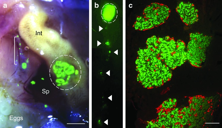
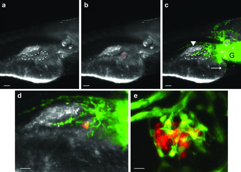
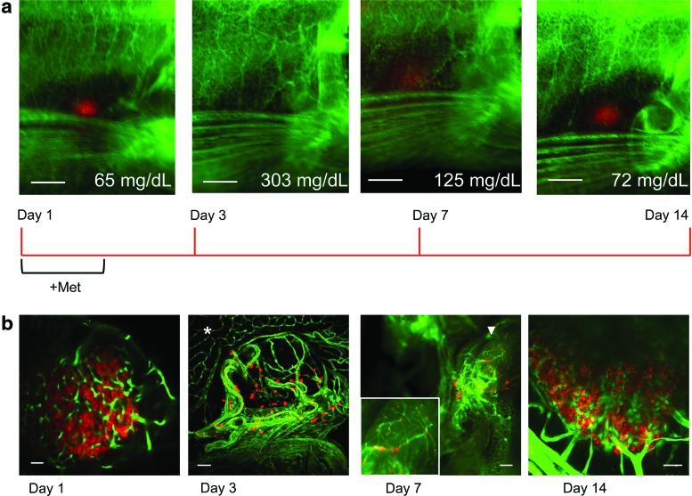
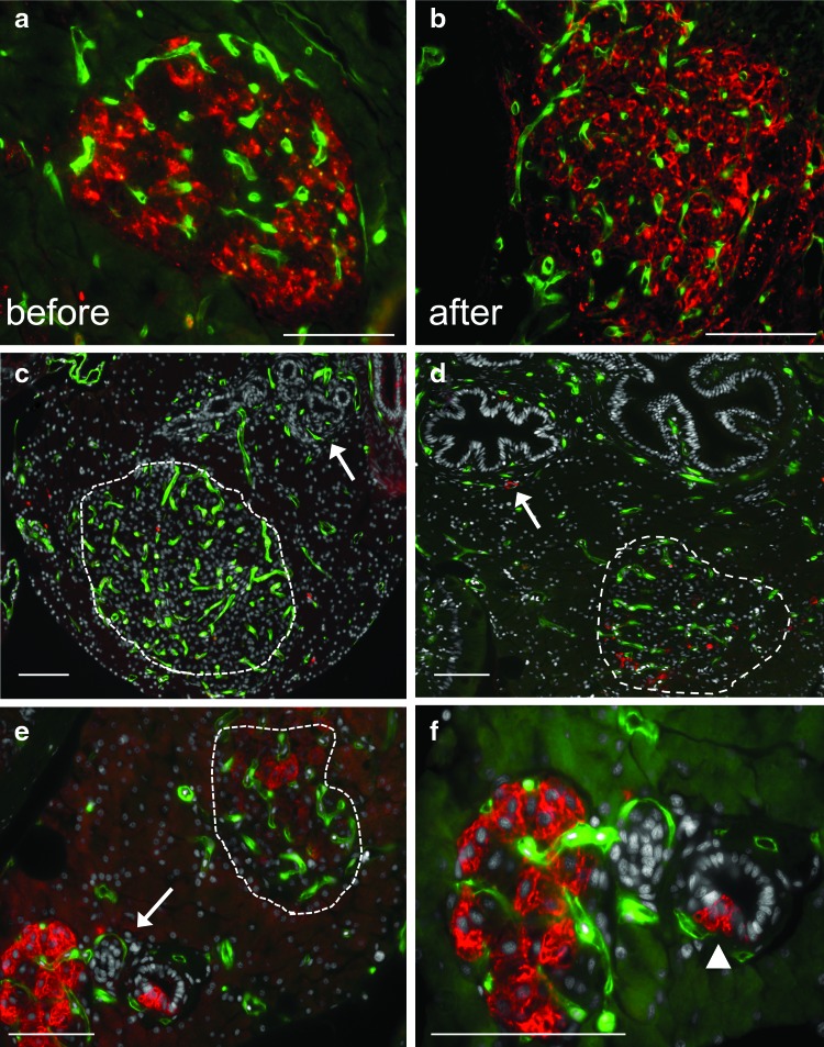
Blood vessel networks provide nutrients and gaseous exchange that are essential for functions. Pancreatic islet capillaries deliver oxygen to endocrine cells while transporting hormones to organs and peripheral locations throughout the body. We have developed a zebrafish diabetes model in which adult islets can be followed in vivo during beta cell regeneration while calibrating changes in beta cell mass and fasting blood glucose levels. After genetic ablation, beta cells are initially dysfunctional or dying, and blood glucose levels increase fourfold. During a 2-week period, hyperglycemia eventually normalizes as beta cell mass regenerates. We show that mCherry-fluorescent, insulin-positive beta cells re-emerge in close contact with the vascular endothelium. Alterations in the dense vascular network of zebrafish islets were visualized by the expression of green fluorescent protein (GFP) in endothelial cells derived from the Fli transcription factor promoter. The rapid destruction and regeneration of beta cell mass was evaluated in the same animal over time, providing a functional model for investigating the interactions of islet cell types with vascular cells as well as the consequences of hyperglycemia on other tissues. Regenerating adult zebrafish can be utilized as vertebrate, metabolically active models for generating new insights into treatments for type 2 diabetes.


Figures

FIG. 1.
FIG. 1.
Multiple islets are organized within the endocrine pancreas of adult zebrafish and express endocrine hormones. (a) Right lateral bright field view of a dissected INSGFP adult female zebrafish with superimposed fluorescent image. Green fluorescent protein (GFP) expression is seen in multiple large islets located in the endocrine pancreas (scale bar=400 μm). Sp: spleen, Int: intestine, L: liver, dashed line: GFP+ islets surrounded by translucent exocrine tissue: bracket. (b) Whole mount fluorescence image of the adult zebrafish endocrine pancreas, right lateral view. Dashed line: multiple large islets. Arrowheads: isolated islets and individual beta cells extending caudally along the intestine. (c) Five micrometer paraffin section of multiple large islets (scale bar=40 μm). Immunohistochemistry was performed without antigen retrieval. Green: endogenous GFP expressed in beta cells within multiple large islets, red: anti-glucagon antibody in alpha cells. Color images available online at www.liebertpub.com/zeb
FIG. 2.
FIG. 2.
The pancreatic vasculature at larval day 6. (a) Bright field image of the right side of a living Insulin Nitroreductase mCherry (INCFli) zebrafish (scale bars in a–c=30 μm). The dashed border defines the pancreas where islet endocrine cells are developing in a single principal islet surrounded by exocrine tissue. (b) Endogenous mCherry fluorescence in transgenic INCFli islets is restricted to beta cells. (c) Endogenous GFP expressed by the Fli promoter within the trunk vasculature. Arrowhead: swim bladder, arrow: heart, G: gills, dashed line: pancreas. (d) Merged red and green fluorescence defines the endocrine pancreas containing nascent blood vessels. Scale bar=60 μm. (e) Multiphoton confocal image of living casper larva. Scale bar=10 μm. Color images available online at www.liebertpub.com/zeb
FIG. 3.
FIG. 3.
Regeneration in living adults, associated changes in blood glucose, and alterations in islet morphology. (a) Images of an affected INCFli adult male casper during regeneration days 1, 3, 7, and 14. Scale bars=0.5 mm. Red: pancreatic beta cells, green: vasculature. The average blood glucose of animals sacrificed at each time point is indicated (mg/dL). A regeneration timeline is depicted below the images. (b) Single multiphoton confocal images of fixed gut sections from affected individuals (1, 3, and 14 day). Scale bar=10 μm. *: vasculature in exocrine pancreas at day 3. Whole mount, Leica MZFlIII microscope image of green vasculature and red beta cells in affected adult on dissection at day 7. Scale bar=100 μm. Arrowhead: multiple large islets. Inset: magnification of multiple large islets. Color images available online at www.liebertpub.com/zeb
FIG. 4.
FIG. 4.
Changes in islet vasculature during beta cell regeneration. Five-micrometer paraffin sections. Representative sections from each time point are shown. Scale bars=20 μm (a–f) endogenous green fluorescence in vasculature. Cell nuclei (stained with Hoechst) are shown in gray (sections c–f). Beta cells are marked with mCherry red fluorescence (a, e, f) or insulin antibodies (b, c, d). (a) Day 1 control (before Met treatment). (b) A regenerated pancreas from an affected INCFli adult sacrificed at day 14 (after). (c, d) Independent, affected male casper transgenics sacrificed at day 3. (c) Arrow points to pancreatic ducts. An islet containing labeled vasculature is circled. (d) A different affected adult sacrificed at day 3 with labeled beta cells in an islet (dashed circle). Arrow points to a beta cell near labeled vasculature surrounding main pancreatic duct. (e) Affected INCFli adult sacrificed at day 7. Arrow points to region magnified in (f) Dashed circle: islet. (f) Regeneration day 7. Magnification of region in (e). Arrowhead: beta cells emerging from small pancreatic duct. Color images available online at www.liebertpub.com/zeb

References

    1. Brissova M. Powers AC. Revascularization of transplanted islets: can it be improved? Diabetes. 2008;57:2269–2271. - PMC - PubMed
    1. Bonner-Weir S. Perspective: postnatal pancreatic beta cell growth. Endocrinology. 2000;141:1926–1929. - PubMed
    1. Rankin MM. Kushner JA. Adaptive beta-cell proliferation is severely restricted with advanced age. Diabetes. 2009;58:1365–1372. - PMC - PubMed
    1. Moss JB. Koustubhan P. Greenman M. Parsons MJ. Walter I. Moss LG. Regeneration of the pancreas in adult zebrafish. Diabetes. 2009;58:1844–1851. - PMC - PubMed
    1. Bouvet M. Hoffman RM. In vivo imaging of pancreatic cancer with fluorescent proteins in mouse models. Methods Mol Biol. 2012;872:51–67. - PubMed

Publication types

MeSH terms

LinkOut - more resources